The American Heart Association (AHA) 2020 Impact Goals aim to increase out-of-hospital cardiac arrest (OHCA) survival from 7.9% to 15% between 2010 and 2020 (Kronick et al, 2015). Both in-hospital and prehospital systems have the potential to substantially improve cardiac arrest care. The European Resuscitation Council (ERC) Guidelines 2010 state that ultrasound might be of diagnostic assistance and guide subsequent treatment of reversible causes (Deakin et al, 2010). The AHA also supports the use of transthoracic echocardiography (TTE) with the condition that it must not interfere with advanced life support (ALS) protocol—particularly chest compressions (Kronick et al, 2015). This can be achieved by performing TTE in the 10-second pulse check window as studies have successfully demonstrated without significant difficulty (Callaway et al, 2015).
Reversible causes with TTE
Massive acute pulmonary emboli (PE) is a reversible cause of cardiac arrest, with thrombolysis being the first line of treatment (British Thoracic Society, 2003). Occlusion of pulmonary blood flow is a common cause of pulseless electrical activity (PEA) arrest and has a reported mortality rate of 20–50% (Zhang, 2017). The British Thoracic Society (2003) guidelines state that TTE will reliably diagnose massive PE. PE caused by an enlarged right ventricle, reduced left ventricular diastolic dimension, decreased ejection fraction slope of the mitral valve, dilated right pulmonary artery, or abnormal septal motion, may be detected with TTE (Torbicki et al, 1992). Additionally, the McConnell sign for acute PE includes a description of normal right ventricular apex contractility with akinesia of the free wall of the right ventricle (Lopez-Candales et al, 2010). Interestingly, McConnell identified that patients with chronic pulmonary hypertension will have abnormal wall motion in all regions; yet in acute PE, the apex function is spared (McConnell et al, 1996). The high negative predictive value (96%) of these results means that any negative result is likely to be true and, therefore, abnormal wall motion in all regions can help to rule out PE (McConnell et al, 1996). TTE can also identify thrombi in the right atrium, right ventricle or major pulmonary artery (Torbicki et al, 1992). If identified in the prehospital environment, early thrombolysis will treat the direct cause, but also increase blood flow to the microvascular circulation of the brain post-arrest (Knowles, 2003). The BTS continues to recommend thrombolysis for massive PE, yet the evidence for improved mortality rates is sparse. In 2014, a meta-analysis of 16 randomised control trials (RCTs) showed that thrombolysis for PE had a lower all-cause mortality rate (95% Confidence Interval (CI), 0.32-0.88) compared with anticoagulant treatment (n=2115) (Chatterjee et al, 2014). Only 1.47% of the patients included in this analysis were categorised as high-risk. Henceforth, this data cannot be extrapolated for thrombolysis in cardiac arrest secondary to PE, and warrants further investigation. It is logical that TTE diagnosis is most accurate in patients with massive acute PE as the TTE changes are most evident if the PE is obstructing more than 30% of the arterial bed (Hernandez et al, 2008). As such, in cases of cardiac arrest secondary to PE, the sensitivity of TTE for these findings will be optimal. Currently, there is no conclusive evidence that TTE improves outcomes in these circumstances, but its potential is being recognised as authors call for further research in this field (Zhang, 2017). Furthermore, initiation of thrombolysis must be in the context of having excluded absolute and relevant risk factors.
When the heart is under tamponade, haemodynamic decompensation occurs as the high intrapericardial pressures prevent the heart from expanding and filling (Vaswani et al, 2016). TTE is a non-invasive investigation that allows the extent of pericardial effusion to be evaluated (Breitkreutz et al, 2010). TTE images will show compression of the right atrium and right ventricle during diastole as a result of extra-mural pressure (Lilly, 2016). Early identification and drainage of the pericardial fluid is the optimal intervention in cardiac arrest caused by tamponade in the non-surgical cardiac cohort (Breitkreutz et al, 2010). Trained physicians are able to perform pericardiocentesis after identifying cardiac tamponade as the cause of arrest with TTE, potentially improving patient chances of survival (Breitkreutz et al, 2010).
Severe hypovolaemia is evidenced by flattened left and right ventricles on TTE, or reduced left ventricular end diastolic area (Hernandez et al, 2008). Diagnosis of hypovolaemia via TTE will prevent harmful therapeutic management; for example, preventing thrombolysis for suspected PE when a ruptured aortic aneurysm is the underlying cause of arrest (Hernandez et al, 2008). Additionally, measurement of the inferior vena cava (IVC) is also of value when identifying the reversible causes. In hypovolaemia, the diameter of the IVC may be <5 mm owing to volume depletion and IVC collapse (Hernandez et al, 2008). This would indicate aggressive fluid administration. Whereas an IVC diameter >20 mm suggests ventricular dysfunction, possibly owing to ischaemia, tamponade or PE (Hernandez et al, 2008). Further to the assessment of IVC diameter, the TTE transducer can be used to identify features of many other life-threatening conditions such as tension pneumothorax, cardiac ischaemia (as views may demonstrate regional contractile dysfunction), and aortic dissection (Robson, 2010). Despite the broadly inconclusive evidence, Zhang (2017) asserts that TTE is able to identify the aetiology of cardiac arrest and treating the reversible causes will save lives.
PEA assessment by TTE
The potential for using TTE is mostly researched in the context of PEA cardiac arrests. By definition, true PEA is the presence of electrical activity as identified on electrocardiogram, and is a phenomenon often seen as a result of a prolonged ALS attempt (Rabiei and Rahimi-Movaghar, 2016). Subsequently, the lack of myocardial motility is visible on TTE. In pseudo PEA however, a significant event has impaired perfusion and commonly impedes cardiac venous return. This means that the ventricles fail to produce an effective contraction despite the continuation of ventricular motion (Rabiei and Rahimi-Movaghar, 2016). It is the latter presentation that is more likely to respond to resuscitation, hence the benefit of differentiating between true PEA and pseudo PEA (Tayal and Kline, 2003; Rabiei and Rahimi-Movaghar, 2016). An in-hospital observational study used TTE on patients who were in PEA or near-PEA with a high index of suspicion for pericardial effusion as the cause (Tayal and Kline, 2003). Of the 12 patients included, eight had pericardial effusion observable on TTE. Of these, seven survived to hospital discharge; three of whom had cardiac tamponade and required emergency pericardiocentesis. The use of TTE also assisted in the detection of capture via transcutaneous pacing in two patients without effusion resulting in an early return of spontaneous circulation (ROSC). Although this study selected patients with a high pre-test probability for pericardial effusion, it demonstrated TTE in the PEA state could lead to better decision-making and outcomes for cardiac arrest patients (Tayal and Kline, 2003). To predict outcomes, a prehospital prospective observational study of non-traumatic adult cardiac arrests aimed to evaluate the benefit of TTE (Aichinger et al, 2012). Out of 42 patients, 40% of those with ventricular activity on initial TTE survived to hospital admission, in comparison with 3.1% survival to admission in those who had cardiac standstill on initial TTE. The sample size of this study was small, with only ten patients presenting with cardiac activity on initial TTE and four surviving (Aichinger et al, 2012). Nonetheless, this proved statistically significant (p=0.008); yet the small sample size of these results limits the application of the predicted outcomes.
There has been just one RCT examining the use of TTE in cardiac arrest to predict outcomes, compared to not using TTE (Chardoli et al, 2012). It showed that TTE could detect causes of PEA but the study had multiple limitations and does not comment on the effect it has on outcomes. The AHA disregarded the conclusion of the RCT owing to the small sample size (n=100) and very high risk of bias (Callaway et al, 2015). Ascertainment bias should be suspected as the results and conclusion are systematically distorted by the knowledge of which intervention the patient is receiving, with no description of efforts to blind (Viera and Bangdiwala, 2007). According to the AHA, the paucity of robust RCTs assessing the benefit of TTE in cardiac arrest continues to limit the evidence base for practice in this field (Callaway et al, 2015).
Prehospital ultrasound
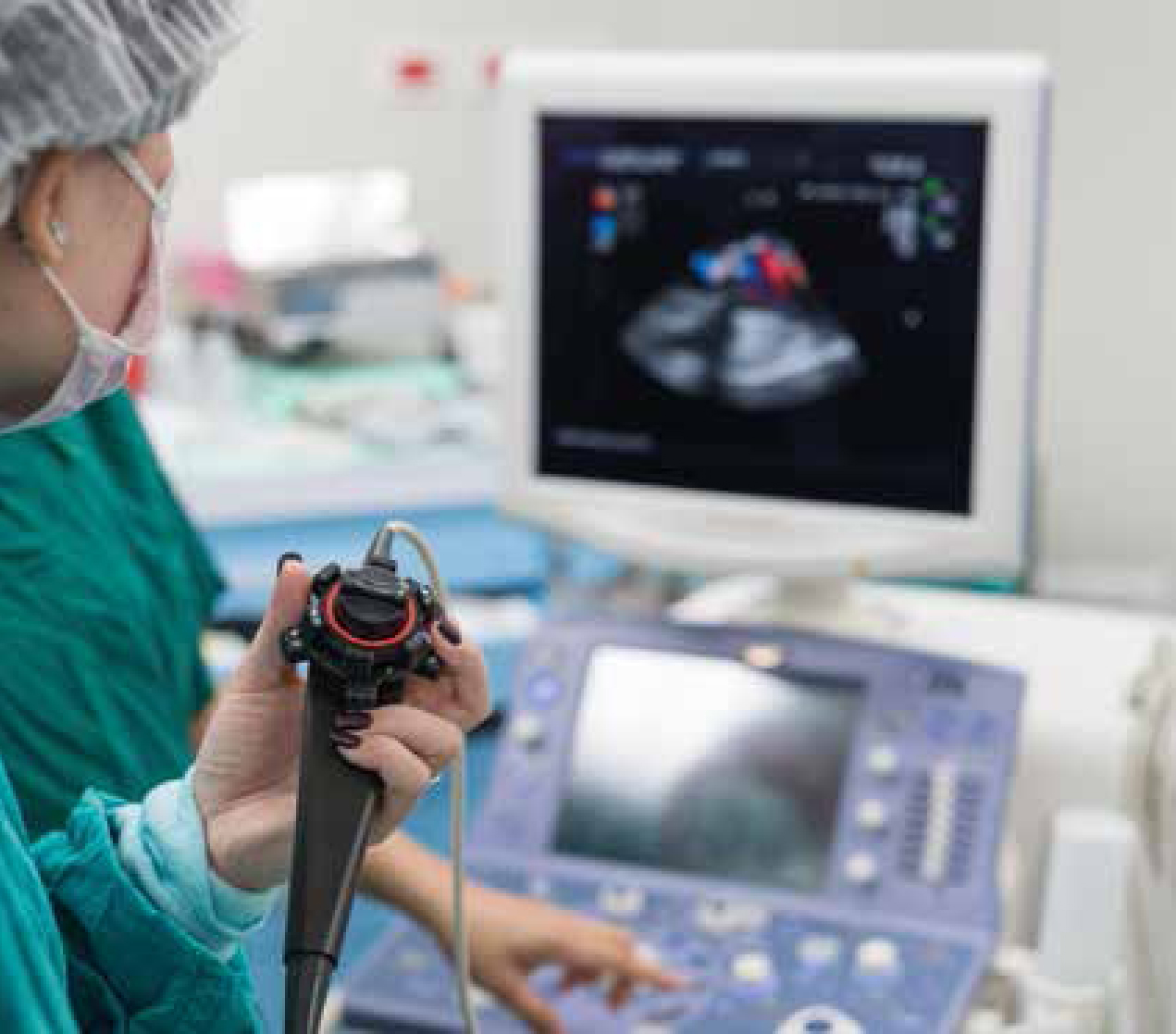
A multitude of international studies have demonstrated the value and feasibility of prehospital ultrasound. The development of portable ultrasound devices has allowed this non-invasive technique to enter the prehospital arena (Busch, 2006). The earlier studies focused on the application of ultrasound in trauma patients only using terms such as FAST (focused assessment sonography for trauma), EFAST (extended FAST) and FASTER (FAST during emergency retrieval). Ultrasound has been shown to reduce the interval time between the onset of pathology and definitive diagnosis in time-critical trauma patients (Busch, 2006). Further studies demonstrated the ease of the technique despite environmental challenges such as performing ultrasound in a moving ambulance or during flight in an aircraft (Mazur et al, 2008; Snaith et al, 2011). Additionally, the quality of prehospital ultrasound images have been shown to equal that of ultrasound images obtained within the emergency department (ED) (Brun et al, 2014). As a result, the application of ultrasound soon broadened to assess medical emergencies, cardiorespiratory compromise and cardiac arrest patients in the prehospital environment (Busch, 2006).
The FAST scan was not developed for implementation in cardiac arrest procedures, nor was it developed for use or interpretation within 10 seconds. Hence the Focused Echocardiography Evaluation in Life Support (FEEL) was introduced to prehospital care. This is an extension of FAST, which allows for quick, limited echocardiography in resuscitation care (Breitkreutz et al, 2010). During resuscitation, the main challenge with FEEL is being able to take adequate images and interpret them within the 10-second pulse check window. It has been demonstrated that trained emergency physicians could perform FEEL in the prehospital setting to alter the management of the cardiac arrest depending on the TTE result (Breitkreutz et al, 2010). This prospective observational study had a modest sample size of 230 patients, yet only 100 were in cardiac arrest. In this arm of the study, adequate subcostal TTE images were obtained in all patients. Coordinated cardiac wall motion was detected in 35% of those with asystole on the ECG and 58% of those with PEA, and associated with increased survival to admission (Breitkreutz et al, 2010). The findings from the TTE directly altered management in 78% of cases. These management alterations included fluid therapy or inotropic support, pericardiocentesis or a change in the choice of hospital destination for specialist intervention. This study demonstrated the feasibility of performing the FEEL examination in the prehospital setting and its value in medical management. ERC guidelines support the use of TTE, yet emphasise the importance of high-quality chest compressions throughout any ALS intervention—pausing only briefly to enable specific interventions such as endotracheal intubation (Deakin et al, 2010).
TTE as a predictor of mortality in out-of-hospital cardiac arrest
TTE can provide clinical information to support the decision to cease cardiopulmonary resuscitation (CPR) in irreversible circumstances, preventing loss of dignity for patients and unnecessary use of valuable resources. There are many other predictors of mortality that have already been well researched, such as end tidal carbon dioxide measurement, the use of early CPR and timely defibrillation. However, studies have shown that TTE findings may provide a more definitive prediction of mortality (Sanders et al, 2001). One retrospective study showed that image-guided resuscitation with TTE helps avoid futile thoracotomy attempts in fatal trauma patients (Ferrada et al, 2014). This was owing to excluding reversible causes, identifying fatal injury and recognising cardiac standstill. A prospective observational study examined the predictive value of cardiac standstill on initial TTE in cardiac arrest patients (Blaivas and Fox, 2001). A complete lack of cardiac activity during a non-traumatic arrest was associated with a 100% mortality rate. To illustrate this, over two-thirds of the patients in this modestly sized study presented with cardiac standstill, none of whom survived to discharge (n=136/169 patients). The authors suggested that this could be an additional indication to cease resuscitative efforts—a factor that would then affect the majority of cardiac arrest patients (Blaivas and Fox, 2001). Salen et al (2005) reached a similar conclusion. A convenience sample of 70 cardiac arrest patients was selected as their initial TTE images showed either asystole or PEA (Salen et al, 2005). Out of 34 patients in PEA, 11 had evidence of cardiac activity (pseudo PEA) and 8 of those achieved ROSC surviving to arrival in intensive care. The accuracy of TTE to predict ROSC versus death in cardiac arrest proved to have a narrow confidence interval (=0.95, 95% CI, 0.92-1.0), which suggests precision in the results (Salen et al, 2005). However, this study does not comment on survival to discharge. Data for long-term survival in all of these studies would be helpful to show the relationship between TTE imaging and subsequent outcome.
Blyth et al (2012) completed a systematic review of TTE as a predictor of survival for in-hospital cardiac arrests. Out of 12 studies involving a total of 568 patients, a lack of cardiac activity of TTE has a significantly lower (but not absent) probability that a ROSC will be achieved compared to those with ventricular wall motion on TTE (Blyth et al, 2012). Blyth et al concluded that cardiac standstill visualised on TTE should not be the sole basis for the decision to stop resuscitation efforts. Extrapolation of in-hospital data has limited clinical application for OHCA yet remains necessary to review due to the paucity of OHCA data.
Introduction of TTE to CCPs
The AHA highlights the difficulties in generalising data from any of the studies above. In the UK alone, there is wide variety of ambulance service organisational structures. Most prehospital TTE studies have been based upon a physician-led structure; yet in the UK, critical care paramedics (CCPs) are more likely to attend an OHCA than a physician. Therefore, introducing TTE to CCPs may be of value in the UK and relevant research is starting to emerge (Walker, 2017).
An observational study in which trained paramedic responders used TTE has shown potential benefit for OHCA patients (Reed et al, 2016). The premise of the study was to perform TTE during the 10-second pulse check window to identify ventricular wall motion. Circulation was restored in 30% of cardiac arrest patients with detectable wall motion on TTE, compared to 2% of patients without wall motion (Reed et al, 2016). There were no changes in management or actions as a result of the TTE findings. However, this study serves as an introduction to paramedic-led TTE within the UK, suggesting that with TTE paramedics may be able to identify cause, predict outcome and alter management accordingly. Arguably, CCP management will remain limited but TTE results may indicate that an emergency physician must be called to the scene to perform advanced skills such as pericardiocentesis or thoracotomy prior to transportation. Equally, if the CCP could identify which intervention is required using TTE, the receiving hospital could prepare appropriate interventions to treat the patient immediately on arrival—further minimising the diagnosis-to-treatment time.
It has been questioned whether non-expert sonographers (physicians and CCPs) are able to obtain high-quality TTE images and subsequently interpret the results (Niendorff et al, 2005). To address this, telemetry of TTE images allows for real-time physician evaluations of the images obtained by physicians or CCPs in the prehospital setting. Multiple authors have demonstrated that telemetry can ensure safe practice by non-expert sonographers. One program in Taiwan used a ‘3G’ communications protocol to transfer TTE images from the ambulance to the receiving hospital ED (Su et al, 2008). This allowed the prehospital clinician to receive instruction to improve care or minimise further harm. Equally, the receiving hospitals were able to prepare for the incoming patient with the relevant surgical teams, equipment and blood.
An American study demonstrated that cardiac-limited ultrasound examination (CLUE), led by cardiologists, was feasible in the prehospital environment (Mai et al, 2013). The study resulted in 100% of successful imaging by novices when guided by a cardiologist via video call from an iPhone. This study also showed that the brevity of the prehospital CLUE was less than 5 minutes, recognising its clinical practicality in emergency situations. These advances in telemetry and video calling may be the safest method of broadening the use of prehospital TTE by CCPs.
Conclusion
The evidence in favour of prehospital ultrasound is undeniable hence various emergency systems worldwide already have the resources and training to perform ultrasound. Non-expert sonographers are able to perform TTE successfully in the prehospital environment; however, the use of telemetry could ensure expert interpretation and patient safety during the introduction of TTE to CCPs in the UK.
A focused TTE assessment has shown to have a role in OHCA in order to evaluate reversible causes with the potential to guide subsequent management. Massive PEs, cardiac tamponade and hypovolaemia can be systematically identified using TTE. Additionally, TTE is a sensitive real-time marker of ventricular activity with the ability to differentiate between PEA and pseudo PEA.
For these reasons, TTE has been shown to better predict mortality rather than survivability in cardiac arrests (Walker, 2017). More research and ethical consideration is required in this area prior to withholding resuscitative efforts in those with cardiac standstill on initial TTE. The impact of TTE results altering OHCA management in the UK has yet to be evaluated. Nevertheless, TTE should form part of the critical care management of OHCA, as its role in reducing time from onset of pathology to diagnosis is indisputable.