Pneumonia is an episode of acute inflammation involving lung tissue and can be of bacterial or viral origin. In the UK, the most common cause of community-acquired pneumonia (CAP) is Streptococcus pneumoniae (Strep. pneumoniae). Strep. pneumonia infections can lead to impaired gaseous exchange, global hypoxaemia, cardiovascular complications, sepsis and death.
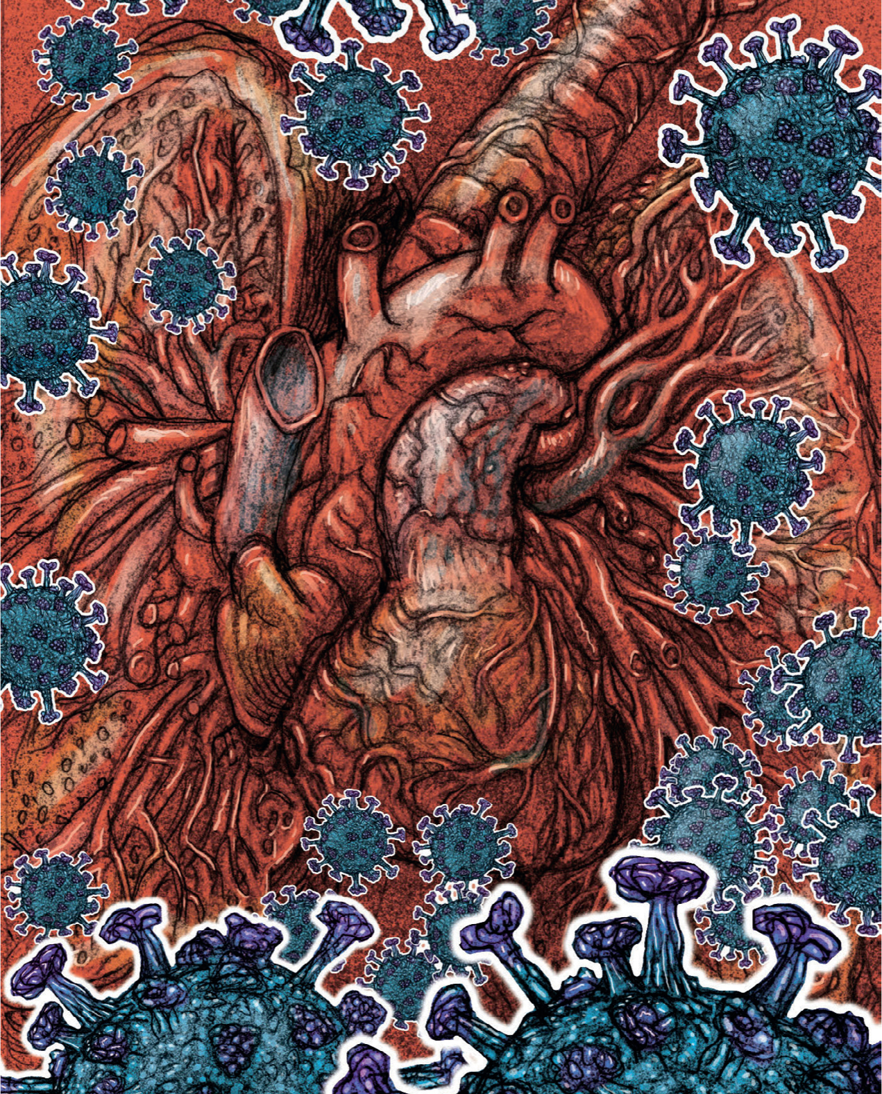
The epidemic of 2019 novel coronavirus (officially called SARS-CoV-2) has expanded from its place of origin, Wuhan in China, to countries around the world. Some of these countries have seen onward transmission (Lipsitch et al, 2020). SARS-CoV-2 is a virus in the zoonotic coronavirus family. This virus, previously unknown in humans, is now causing the disease commonly referred to as COVID-19. Zoonotic diseases are infectious diseases that have spread from non-human animals to humans (Torok, 2016).
Coronaviruses cause respiratory and intestinal infections in animals and humans (Cui et al, 2019) but were not thought to be highly pathogenic to humans until the outbreak of severe acute respiratory syndrome (SARS) in 2002 and 2003 in Guangdong province in China (Cui et al, 2019). Like other coronaviruses, SARS-CoV-2 uses the angiotensin-converting enzyme 2 (ACE2) as a receptor to gain entry into cells, and subsequently infects bronchial epithelial cells and lung cells called type II pneumocytes (Qian et al, 2013; Boniotti et al, 2016; Saif et al, 2019).
Signs and symptoms of SARS-CoV-2 include a cough, pyrexia (above 38°C) and shortness of breath. Additional symptoms may include muscle aches, lethargy, a runny nose and tiredness (NHS, 2020). A small percentage of infected patients may go on to develop severe bilateral pneumonia and multiple organ failure, and die.
Normal anatomy and physiology of the lungs
The lungs have multiple protective mechanisms to prevent lower respiratory tract infections such as pneumonia. The curved, undulating nature of the upper airways creates a great deal of turbulence in air as it travels through these airways. This turbulence allows the hairs and mucus that line the airways to trap inhaled particles, so particles approximately the size of red blood cells will not enter the lung (Hall, 2011). The sneeze reflex acts to eliminate inhaled particles; this is initiated by irritation of the nasal passages, and the resulting rapid movement of air through the nasal passages during sneezing expels the irritative substance.

The walls of the trachea are lined with goblet cells, which produce a protein-rich mucus that has antibacterial peptides so it destroys microbes as they become trapped in the mucus layer. In addition, the trachea is lined with cilia, which have the ability to move in a beat-like fashion (Lumb, 2011). This movement transports mucus laden with inhaled particles upwards to the larynx, where it is either swallowed or expectorated via the cough reflex (Patton and Thibodeau, 2013).
These defence mechanisms will keep the majority of foreign bacteria out of the alveoli and reduce the risk of infection. However, very small particles—such as those found in cigarette smoke, or industrial dust such as slate or coal—do find their way to the alveoli. This is because the sheer volume of the inhaled particles surpasses the defence mechanism's ability to entrap and eliminate them. Alternatively, rapid breathing means that even large particles remain suspended in the inhaled air and are transported to the alveoli (Lumb, 2011).
Defence mechanisms within the alveoli include immune cells such as macrophages, which identify and digest (phagocytose) invading pathogens. Migration of these macrophages towards a pathogen is controlled by a surfactant protein that also stimulates the production of antibodies to eliminate invading pathogens (Lumb, 2011). The surfactant is a four-protein substance produced by type II alveolar cells. Two of the surfactant proteins act to reduce surface tension in the alveoli, which maintains alveolar expansion and facilitates gas exchange. The remaining two surfactant proteins stimulate immune responses within the alveoli to eliminate invading pathogens.
Both alveolar macrophages and surfactant proteins can usually reduce the risk of infection and help maintain the integrity of the alveolar membrane. This alveolar membrane is the site of gas exchange between the alveoli and pulmonary circulation, and has an approximate surface area of 70 m2 that is essential to maintain appropriate rates of oxygen transport to pulmonary blood and eliminate carbon dioxide (Tortora and Derrickson, 2011). This alveolar membrane is closely surrounded by a mesh of pulmonary capillaries and this degree of proximity is essential in maintaining effective diffusion of gases across the alveolar membrane (Lumb, 2011).
Effective gas exchange relies upon ventilation (V), which is the inspiration of gases into the alveoli, and perfusion (Q), which is the blood flowing through the pulmonary circulation. This relationship is referred to as V/Q and a compromise in either V or Q will result in reduced gas exchange. For example, if a patient experiences a pulmonary embolism, the perfusion to the area distal to the embolism will not participate in gas exchange, and the area effectively becomes dead space, resulting in hypoxia and hypercapnia. If an area is poorly ventilated, which happens in pneumonia, perfusion to that area will be reduced as the pulmonary circulation vasoconstricts (in areas of hypoxia), effectively shunting blood to other, better ventilated areas. This is explored in more detail in relation to pneumonia below.
Pathophysiology of pneumonia
Pneumonia is an episode of acute inflammation involving lung tissue and can be of bacterial or viral origin. Pneumonia that affects multiple areas of the lung is termed bronchopneumonia, while that affecting a single lung lobe is known as lobular pneumonia (Lumb, 2011). The type of bacteria or virus that causes the pneumonia will usually depend on whether the infection is community-acquired pneumonia (CAP) or hospital-acquired pneumonia (HAP), with immunocompromised patients usually succumbing to other pathogens (Table 1).
| Community-acquired pneumonia | Hospital-acquired pneumonia | Immunocompromised |
|---|---|---|
| Streptoccocus pneumoniae | Pseudomonas aeruginosa | Pneumocystis jirovecii |
| Haemophilus influenzae | Staphylococcus aureus | Mycobacterium tuberculosis |
| Staphylococcus aureus | Klebsiella pneumoniae | Atypical mycobacteria |
| Mycoplasma pneumoniae | Enterobacter | Respiratory viruses |
| Chlamydophila pneumoniae | Protozoa | |
| Moraxella catarrhalis | Parasites | |
| Streptococcus pneumoniae | ||
| Influenza | ||
| Rhinovirus | ||
| Coronavirus |
Paramedics will be familiar with attending calls to patients who have developed CAP, which is most commonly caused by Strep. pneumoniae, so this article will focus mostly on this scenario. However, the pathophysiological principles are similar for all bacterial and viral causes.
When a pathogen reaches the alveoli, a series of inflammatory responses involving alveolar macrophages and surfactant proteins occur. Their effects include inflammation of the respiratory membrane and migration of neutrophils from the pulmonary circulation into the lung tissue (Lumb, 2011). This is achieved through the release of numerous inflammatory mediators, which make neutrophils stick to the endothelial cells that line the pulmonary capillaries; these also cause the neutrophils to drift apart, which allows them to migrate between the cells. These neutrophils play a vital part in the destruction of invading pathogens by producing lysosomes containing enzymes that break down pathogens (McCance and Huether, 2014).
Fluid and plasma proteins simultaneously migrate with the neutrophils causing flooding of the affected alveoli with fluid known as exudate (Patton and Thibodeau, 2013). Furthermore, the inflammatory mediators damage the alveolar membrane that usually maintains gas exchange.
These two processes result in an increase in the distance between the alveolar membrane and the pulmonary capillaries, and also reduce the functional surface area for gas exchange, causing hypoxia with a compensatory increase in respiratory rate.
Furthermore, the presence of the pathogen in the alveoli reduces surfactant activity, resulting in higher surface tension within the alveoli and therefore alveolar collapse (atelactasis), again reducing the gas exchange capacity of the lung (Hall, 2011).
The damaging consequences of the infection and inflammatory process adversely impact ventilation to the affected area causing a shunt, whereby pulmonary vasoconstriction causes blood to be moved to better ventilated areas of the lung, which results in a V/Q mismatch (Lumb, 2011).
This initial stage results in areas of lung becoming consolidated; affected areas have a distinct red appearance as a result of the oedematous fluid and blood cells gathering in the area, which gives it the name red hepatisation. As neutrophil numbers increase in the area and fibrin is deposited in the lung tissue, the lung turns a grey colour, which explains its descriptor of grey hepatisation (McCance and Huether, 2014).
Pathophysiology of COVID-19 pneumonia
Like other respiratory pathogens, including influenza and rhinovirus, the novel coronavirus spreads primarily through droplets of saliva or discharge from the nose when an infected person coughs or sneezes (World Health Organization (WHO), 2020a). According to Guan et al (2020), the incubation period for COVID-19 is thought to be within 14 days of exposure. However, most cases present approximately 4–5 days after exposure.
Recent reports suggest that COVID-19 is associated with severe disease that requires intensive care in approximately 5% of proven infections (Wu and McGoogan, 2020). Additional evidence suggests that pneumonia is the most frequent serious manifestation of this COVID-19 infection (Guan et al, 2020; Lee at el, 2020). Cascella et al (2020) maintain that the pathogenic mechanism that produces COVID-19 associated pneumonia seems to be particularly complex. The long incubation period made it difficult to prevent transmission during the early days of the current outbreak, as many health workers in Wuhan became infected when they were seeing patients without sufficient protection, according to Tian et al (2020).
At present, there is a limited amount of pathologic data on this novel coronavirus-associated pneumonia from either autopsy or biopsy. However, evidence demonstrates that the coronavirus infects bronchial epithelial cells and type II pneumocytes, using ACE2 as a receptor to gain entry into cells (Qian et al, 2013; Boniotti et al, 2016; Saif et al, 2019). According to Lee et al (2020), the predominant CT findings of patients with COVID-19 include ground-glass opacification, consolidation, bilateral involvement, and peripheral and diffuse distribution. These findings concur with those of other reports in smaller cohorts (Chung et al, 2020; Lee et al, 2020). According to Tian et al (2020), the lung parenchyma of patients with COVID-19 appears patchy, with evident proteinaceous and fibrin exudates. There is also diffuse thickening of alveolar walls, with proliferating interstitial fibroblasts and type II pneumocyte hyperplasia. These lung changes severely affect oxygen diffusion, leading to global hypoxia (WHO, 2020b).
According to Cascella et al (2020):
‘The COVID-19 may present with mild, moderate, or severe illness. Among the severe clinical manifestations, there are severe pneumonia, ARDS [acute respiratory distress syndrome], sepsis, and septic shock. The clinical course of the disease seems to predict a favourable trend in the majority of patients. In a percentage still to be defined of cases, after about a week there is a sudden worsening of clinical conditions with rapidly worsening respiratory failure and MOD/MOF [multiple organ dysfunction/multiple organ failure]. As a reference, the criteria of the severity of respiratory insufficiency and the diagnostic criteria of sepsis and septic shock can be used.’
According to the WHO (2020a; 2020b) approximately 14% of patients with COVID-19 develop severe disease that requires hospitalisation and oxygen support, and 5% require admission to an intensive care unit (Wu and McGoogan, 2020). In severe cases, COVID-19 can be complicated by acute respiratory distress syndrome (ARDS), sepsis and septic shock, multiorgan failure, as well as acute kidney injury and cardiac injury (WHO, 2020a). Older age and comorbid disease have been reported as risk factors for death.
Risk factors and causes
Pneumonia develops because of a compromise in an individual's defence systems; this could be a result of other illnesses or another pathophysiological process. The immune response becomes less acute, increasing susceptibility to infection. Furthermore, alveolar macrophage activity decreases, as does that of the ciliary system, which explains why the majority of those who develop pneumonia are aged over 65 years—as old age and comorbidities lead to reduced alveolar macrophages (Tortora and Derrickson, 2011). Patients with a diminished gag reflex are also at risk of developing pneumonia; these could be individuals who have experienced a cerebrovascular accident in the past or sustained a head injury (Banasik and Copstead, 2013).
Paramedics will probably be familiar with attending calls to patients with long-term conditions such as chronic obstructive pulmonary disease (COPD). These patients, who tend to be chronically hypoxic, have multiple risk factors for pneumonia, such as smoking and chronic lung inflammation, which make them immunocompromised (Dahl and Nordestgaard, 2009). Additionally, optimal functioning of the immune system requires adequate levels of oxygenation, and a COPD patient's chronically hypoxic state blunts the immune response, thus increasing their risk of developing pneumonia (Hall, 2011). Box 1 outlines the risk factors for developing CAP.
Clinical presentation
Common clinical symptoms seen in pneumonia generally involve fever and chills indicative of an infection, but the presence of a productive cough can vary, depending on the causative pathogen (Banasik and Copstead, 2013). However, the cough will become more productive in the latter stages of an infection (Lumb, 2011), with Strep. pneumoniae tending to present with a productive cough with rust-coloured sputum. Creed and Spiers (2010) state that pneumonic patients will also be breathless and complain of pleuritic chest pain that worsens on inspiration and coughing. This sustained period of breathlessness and the high probability that the patient will be elderly mean that the risk of the patient tiring and developing respiratory failure is all too real and should be considered.
Because of the high risk of pneumonia becoming a systemic infection, the paramedic should observe for signs of systemic inflammation, including tachypnoea, hypotension, tachycardia, hypoxia and confusion (Creed and Spiers, 2010).
Paramedics should bear in mind when assessing a patient that only some of the above clinical symptoms may be evident and also that they may be mild in their presentation. As with any acute illness, early identification and intervention are key to maximising patient outcome, so knowledge of risk factors is essential in maintaining a high index of suspicion surrounding pneumonia.
Assessment and management of COVID-19
The complex nature and the non-specific signs and symptoms of COVID-19 make the management of affected patients challenging. From the multitudes of cases being identified daily, more signs and symptoms are being added for potentially infected patients, although some of the evidence for this is ad hoc and unsubstantiated. Examples of these include conjunctivitis (1–3% of cases) and early data suggest anosmia and ageusia (lack of smell and taste).
Before any patient assessment can take place, appropriate personal protective equipment should be readily available.
A detailed history should be undertaken as this can be used to diagnose 80–85% of cases (Innes et al, 2018). This depends on how critically stable the patient is but, again, because of the vagueness of COVID-19, a diagnosis may not always be reached. A high index of suspicion for COVID-19 must be considered when presented with any of these signs and symptoms.
For any patient, the ABCDE approach should be adopted as this enables any life-threatening conditions to be identified at the earliest opportunity and treated in that order (Resuscitation Council (UK), 2015).
A simple look, listen and feel approach should be instigated to assess the airway to determine if there are immediate changes that need remedying. COVID-19 is not generally associated with airway compromise. Following the airway assessment, a comprehensive assessment of breathing will follow. This includes looking at the respiratory rate, the effort of breathing, the use of accessory muscles and any noises that are audible without the use of a stethoscope (Johnson et al, 2015). The paramedic should be familiar with and proficient in undertaking a respiratory assessment, including auscultation and percussion, and understand adventitious breath sounds, percussion notes and the associated pathophysiology; this is of paramount importance (Curtis and Ramsden, 2015). COVID-19 may present in a similar way to pneumonia, with crackles present (these are abnormal lung sounds characterised by discontinuous clicking or rattling sounds) and percussion of the lung fields can be carried out to identify hyporesonance, which indicates fluid (Ruthven, 2016). These could indicate ARDS, a serious, life-threatening complication of COVID-19. ARDS needs to be managed in an intensive care setting so early identification is key for patient survival.
As part of an assessment for hypoxaemia and oxygen requirements, the British Thoracic Society (BTS) (2015) recommends the use of pulse oximeters. Pulse oximeters allow for simple assessment of oxygenation and should be available in all locations where emergency oxygen is used. Peate (2014) maintains that the ability of the paramedic to monitor oxygen levels using a pulse oximeter is key to high-quality, effective prehospital care and it is important to use oximeters for assessing ambulatory patients with suspected COVID-19. If blood saturation is low, supplementary oxygen should be administered and oxygen delivery should aim for an oxygen saturation level (SpO2) of 94–98%. (BTS, 2015). Paramedics can treat pulmonary oedema of cardiac origin but cannot treat pulmonary oedema associated with ARDS.

A circulatory assessment should include palpation of pulses, measurement of blood pressure and recording an electrocardiogram (ECG) as a minimum and, if trained to do so, paramedics can also assess jugular venous pressure and auscultate heart sounds (Curtis and Ramsden, 2016). Following these assessments, the paramedic can treat underlying issues identified such as any arrhythmias or hypotension. Any treatment given should be in line with current guidelines such as those by the National Institute for Health and Care Excellence (NICE), Joint Royal Colleges Ambulance Liaison Committee (JRCALC) or Scottish Intercollegiate Guidelines Network (SIGN).
The disability element of the ABCDE assessment assesses the neurological status of the patient. An initial history-taking with the patient gives a good sense of the level of cognition. It is important to establish a baseline Glasgow Coma Scale score, plasma glucose, pupillary response and temperature before any patient deterioration (Gregory and Ward, 2014). Any deficiencies noted in plasma glucose can be addressed rapidly. Current JRCALC clinical guidelines from the Association of Ambulance Chief Executives (AACE) (2019) do not recommend the use of paracetamol to treat pyrexia alone unless it is associated with discomfort. Finally, if appropriate, a full head-to-toe assessment looking for any skin changes, rashes or wounds that could be the source of any infection needs to be performed (Pilbery and Lethbridge, 2019).
Once observations have been collated, they can be added to the National Early Warning Score (NEWS/NEWS 2) chart. This tool can aid the paramedic in identifying the deteriorating patient and providing receiving units with a standardised score; note, however, this tool does not replace clinical judgement (Royal College of Physicians, 2017).
Conclusion
Pneumonia is an infectious process resulting from the invasion and overgrowth of microorganisms in lung parenchyma, most commonly by Strep. pneumoniae. This can lead to the development of intra-alveolar exudate and consolidation. If it is not treated early, multiple respiratory, cardiovascular and inflammatory complications can occur.
Paramedics can have a profound impact on the care of patients with pneumonia. The effective management of the disease by the paramedic involves prompt recognition and early administration of oxygen, intravenous fluids, antibiotics and transfer to hospital.

