In a prehospital setting, the ‘foot of the bed inspection’ becomes an ‘over the ambulance dashboard inspection’. A mangled wreck at the foot of a tree is usually a good indication that someone has been injured, although mechanism alone has not been proven to be an accurate predictor of trauma severity (Boyle, 2007). A systematic approach incorporating both the mechanism of injury and physical assessment reinforces the index of suspicion and helps determine whether or not the patient is time critical.
The introduction of ‘Golden Hour’ and ‘Platinum Ten Minutes’ has arbitrarily and unwittingly contributed to a target driven prehospital environment that is arguably governed by the clock and not the clinician. Now, in the midst of NHS reconfiguration, hospitals come trauma centres are set to become bigger and better— and much further apart.
Longer transfer times will mean that the ‘scoop and run’ option will not necessarily be the best course of action for seriously ill and injured patients—paramedics and doctors simply have to get better at looking after them in the field. As paramedics’ assessment skills improve and doctors gain prehospital experience, it is anticipated that a well balanced team will emerge—a team that is aware of their limitations and limit their interventions to the time permitted to intervene.
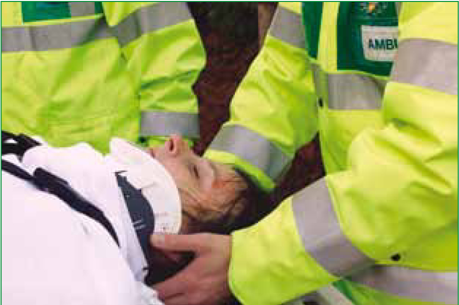
The case study that follows is based on the young male driver of a vehicle that has been involved in a high speed collision with a tree. The patient was unrestrained and ejected from the vehicle on impact. When the author arrived on scene he had already been immobilized on a spinal board. The aims of this case study are to identify the probable pathologies, explain the pathophysiology of the clinical signs and discuss, with evidence, the treatment options and appropriate destination for the patient. It is worth noting that this study focuses mainly on the primary survey.
Airway
Although the patient's airway is patent on arrival, it is important to anticipate difficulties should the patient's condition deteriorate. A patient that is supine on a backboard is vulnerable should they vomit. Vomiting is triggered by a variety of mechanisms including mucosal irritation, stomach distension and stimulation of the chemoreceptor trigger zone by certain drugs (Ganong, 2005; Tortora and Grabowski, 2006). The clinical syndrome of shock causes sympathetic vasoconstriction that limits blood flow to the abdominal viscera—this reduces gastric motility and increases the chance of vomiting (Ganong, 2005). Alcohol may cause gastric irritation; the circumstances surrounding this incident may indicate alcohol intoxication.
It is also important to consider the side-effects of certain analgesic agents. The chemoreceptor trigger zone is a circumventricular structure and, as such lacks the protection of the blood-brain barrier. Opioid analagesics, such as morphine, can therefore stimulate the chemoreceptor trigger zone and cause vomiting (Tortora and Grabowski, 2006). Taking all the above airway risk factors into account, it is essential to allocate airway management to a competent clinician to pre-empt and avoid catastrophic airway compromise.
Breathing
In the prehospital environment, ambient noise can make detailed chest examination difficult, masking the subtleties of potentially life-threatening chest injuries. In this case however, the patient is clearly in respiratory distress. ‘A high, or increasing, respiratory rate is a marker of illness and a warning that the patient may deteriorate suddenly’ (Resuscitation Council (UK), 2006: 9) and although the rate of deterioration can vary (Leigh-Smith and Harris, 2005), tachycardia and tachypnoea following rapid, unrestrained deceleration should be considered a pre-terminal sign until proven otherwise.
Intrapleural pressure (IPP) in the normal lung is subatmospheric and ranges from -2.5 mmHg to -6 mmHg during quiet inspiration and as low as -30 mmHg during forced inspiration. Any disruption to either the lung or chest wall that permits direct communication between the pleural space and the atmosphere causes equilibrium between atmospheric and intrapleural pressures on the affected side. Air in the pleural space is called a pneumothorax. In rare cases, a flap of tissue over the insult can act as a one way valve, permitting air into, but not out of the pleural space (Ganong, 2005). This is called a tension pneumothorax and causes intrapleural pressure to rise above atmospheric pressure ‘leading to lung collapse, chest wall expansion, diaphragmatic depression and contralateral lung compression’ (Leigh-Smith and Harris, 2005: 9).
A tension pneumothorax is a time critical medical emergency. Common signs and symptoms are chest pain, respiratory distress and tachycardia. Due to the maximal inspiratory effort required to overcome increasing IPP, ipsilateral hyper-expansion occurs (Leigh-Smith and Harris, 2005) and decreased air entry is noted in 50-75% of cases (Lee et al. 2007) on the affected side. Tracheal deviation is a late sign and unlikely to be seen in conscious patients. The Joint Royal College Ambulance Liaison Comittee (JRCALC) Guidelines (2006) states that patients are usually hypotensive as a result of decreased cardiac output, however this is uncommon in awake patients. This could be due to awake patients lacking the capacity to produce sufficient IPP to cause great vessel obstruction (Leigh-Smith and Harris, 2005).
Once confirmed, tension pneumothorax must be decompressed by needle thoracocentesis while administering high flow oxygen through a non-rebreathing mask (JRCALC, 2006). The efficacy and appropriate use of this procedure has been questioned, but there is little doubt that a tension pneumothorax can prove fatal if not treated promptly (Fitzgerald et al, 2008). Pulse oximetry and ECG monitoring should be used to assess hypoxia. In the author's experience, paramedics have always been trained to perform thoracocentesis using anterior landmarks (2nd intercostal space in mid-clavicular line) although JRCALC (2006) do not specify. It is suggested that lateral decompression (5th intercostal space in anterior axillary line) may provide a safer alternative as it avoids major vessels and lung tissue that lie closer to the anterior chest wall (Rawlins et al, 2003). In the presence of SpO2 <90% (on high O2), a respiratory rate of <10 or >30 or inadequate chest expansion, assisted ventilation should be considered (JRCALC, 2006).
Once needle thoracocentesis has been performed, it is essential to regularly reassess the patient's chest for bi-lateral air entry, chest movement and normoresonance until a definitive chest drain has been inserted (Laws et al. 2003).
Circulation
Blood loss due to trauma can be internal or external and, if allowed to persist, can cause hypovolaemic shock. Pallor, cool peripheries, tachycardia and absence of palpable radial pulses are a clear sign that sympathetic output has been increased and significant haemorrhage should be suspected (Ganong, 2005; JRCALC, 2006). In this case, the patient is complaining of bi-lateral groin pain and has external rotation to both legs indicating possible pelvic fractures. The presence of a painful, swollen right thigh is suggestive of a fractured femur.
Haemorrhage into the pelvic cavity following trauma has the capacity to far exceed available circulating volume (Grimm et al. 1998) and closed fractured femurs can contain up to 1500 ml of blood (Lee and Porter, 2005; Stahel et al. 2005). The patient is also complaining of lower abdominal tenderness when palpated. This may be directly related to the pelvic injury, but taking the high mechanism and other injuries into account, serious abdominal injury should be suspected until proven otherwise.
Hypovolaemic shock is characterized by reduced cardiac output due to an inadequate circulating volume of vascular fluid (Ganong, 2005). If haemorrhage is controlled, homeostatic mechanisms can compensate for up to 10% blood loss by means of the sympathetic nervous system (Tortora and Grabowski, 2006). Peripheral vasoconstriction is intensified by the release of adrenaline and noradrenaline which increases systemic vascular resistance; this increases the heart rate (chronotropic) and force (inotropic) of contraction and helps to maintain blood pressure. If however the bleeding continues, the compensatory mechanisms begin to fail.
Once the mean blood pressure falls below 60 mmHg, coronary perfusion pressure drops causing myocardial ischaemia, resulting in reduced cardiac output and the inevitable deterioration of the patient (Tortora and Grabowski, 2006).
Provided that the patient's airway is being managed appropriately, attention should now focus on arresting internal haemorrhage and fluid replacement therapy. JRCALC (2006) prioritize haemorrhage control over fluid replacement but in this case, appropriate tasking of available staff should allow simultaneous treatment. While maintaining oxygen delivery of 15 l/ min, consideration should be given to traction and splintage of fractures. Lee and Porter (2007) state, with regard to pelvic fractures, that ‘with circumferential pressure and stabilization, bony bleeding is reduced by apposition of the fracture site, and the reduced movement of bone ends prevents disruption of a formed clot’ (2007:132).
In the prehospital setting, pelvic fractures can be stabilized using triangular bandages, Kendrick Extrication Device (KED) or pelvic straps. Springing the pelvis to confirm the presence of a fracture is now believed to be unreliable and potentially dangerous with regard to dislodging clots and increasing blood loss (Lee and Porter, 2007).
In closed femur fractures, blood loss can be exacerbated by bony overlap and open venous channels. By applying traction, the bone is realigned, venous channels closed and the potential space for bleeding is reduced (Lee and Porter, 2005). Traction can also reduce pain, help prevent fat embolism and minimize the risk of opening a closed fracture (JRCALC, 2006).
It is important to remember that prehospital traction is applied using the pelvis as a static point (JRCALC, 2006), therefore the risk of increasing pelvic injury should be considered against the benefits of applying traction to a mid shaft femur. Injuries should always be assessed and continually reassessed following immobilization to minimize the risk of neurovascular compromise.
‘If all seriously ill and injured patients are to get the best chance of survival then paramedics and trauma centres must evolve together.’
Fluid replacement
Reviews of prehospital fluid replacement in trauma have provided little, if any, evidence that the practice is beneficial and suggest that it may actually do more harm than good (Dretzke et al, 2006). The rationale behind fluid replacement is to ‘reverse the effects of hypovolaemia by increasing circulatory blood volume and blood pressure back towards normal, in order to maintain the perfusion of vital organs and to reduce the risk of death from multiple organ failure’ (UK National Institute for Clinical Excellence, 2004: 6). Conversely, aggressive fluid replacement to normal physiological levels may cause clot disruption and dilutional coagulopathy, leading to exacerbation of haemorrhage (McDonald and Ryland, 2008).
Taking this into account, the National Institute for Clinical Excellence (NICE) guidelines (2004) suggest that fluid replacement is indicated by the absence of a palpable radial pulse or, in the case of penetrating torso injuries, the absence of a central pulse. An initial 250 ml bolus of crystalloid should be administered via a large bore cannula, with a view to obtaining and maintaining either a radial or central pulse in accordance with the patient's injuries. This may be continued to a maximum of 2 litres (JRCALC, 2006).
Analgesia
Once immediately life-threatening conditions have been addressed, it is important to provide adequate and effective pain relief. Failure to do so is unethical, may complicate patient assessment and can have a detrimental effect on physiology (Zohar et al, 2001; Ricard-Hibon et al, 2008). The current JRCALC guidelines advocate the use of two analgesic agents for severe pain; Entonox® and morphine sulphate. Entonox® is an inhaled analgesic gas that must be self administered—this makes it impractical for combative or neurologically impaired patients. Furthermore, JRCALC (2006) recommend cautious administration of Entonox® for patients with suspected pneumothorax, in the absence of a chest drain, due to the risk of increasing intrapleural pressure. Morphine sulphate is a strong opioid analgesic and although JRCALC (2006) consider it to be the ‘gold standard’ for parenteral administration, there are important side-effects to consider.
As previously mentioned, morphine can cause nausea and vomiting and this can seriously compromise patient management. Metoclopramide can be administered to counteract these effects but can induce acute dystonic reactions, especially in children and young adults (Joint Formulary Commitee, 2010). Morphine is also associated with respiratory depression and hypotension. JRCALC (2006) state that it should not be administered to adult patients with a systolic blood pressure less than 90 mmHg and that in the presence of haemorrhagic shock, it may precipitate irreversible hypotension. Prior to fluid replacement therapy, the patient did not have palpable radial pulses and as such, it is possible that morphine is contra-indicated. A radial pulse, however, is not a reliable measure of systolic blood pressure (Deakin and Low, 2000)
Several studies have suggested that ketamine should be more readily available as an analgesic in the prehospital setting. Sympathomimetic effects help to maintain blood pressure, even in hypotensive patients (Svenson and Abernathy, 2007); spontaneous respiration, airway tone and reflexes are usually maintained. Ketamine also promotes gut motility, so post administration vomiting is unusual (Wood, 2003).
Disability
In the prehospital setting, conscious level is measured by using the Glasgow Coma Score (GCS) (Box 1) or the AVPU scale (Alert, Verbal, Pain, Unresponsive). In this case, the patient is ‘drowsy’, responds to voice by opening his eyes and replies with confused speech; he obeys by moving his arms and wiggling his toes. With a GCS 13 and a ‘Verbal’ response on the AVPU scale, it can be said that he has a reduced level of consciousness. Many factors affect a patient's level of consciousness— such as hypoxia, hypovolaemia, hypoglycaemia.
| Item | Score |
|---|---|
| Eyes opening: | |
| Spontaneously | 4 |
| To speech | 3 |
| To pain | 2 |
| None | 1 |
| Motor response: | |
| Obeys commands | 6 |
| Localizes pain | 5 |
| Withdraws from pain | 4 |
| Abnormal flexion | 3 |
| Extensor response | 2 |
| No response to pain | 1 |
| Verbal response: | |
| Orientated | 5 |
| Confused | 4 |
| Inappropriate words | 3 |
| Incomprehensible sounds | 2 |
| No verbal response | 1 |
Hypoxia is a state of inadequate oxygen delivery at a tissue level and can be caused, or exacerbated by hypovolaemia (Ganong, 2005). A consequence of hypoxia is cerebral ischaemia, which can cause mental aberration comparable to that of alcohol consumption (Ganong, 2005). This may belie injury severity and as such, delay the treatment of time critical pathology.
Hypothermia is also a common complication of polytrauma with symptoms not dissimilar to alcohol intoxication. It is worth noting that hypothermia may mask hypoglycaemia (Greaves et al, 2006). It is therefore important to objectively examine all patients with a decreased level of consciousness. The slightest indication that someone has been drinking may be a very misleading sign. Ongoing oxygen and fluid therapy may be indicated to correct hypoxia and hypovolaemia. The patient's pupils are size 4, equal and reactive. It is essential to monitor GCS and pupillary signs closely for prompt detection of developing brain pathology (Cowley and Da Silva, 2008).
Exposure and hypothermia
Hypothermia is a common complication of polytrauma and is considered to be present when a patient's core temperature is below 35 ˚C (McDonald and Ryland, 2008). It is characterized by shivering (above 33 ˚C), confusion, vasoconstriction, etc. Shivering depletes the muscle stores of glycogen and increases oxygen consumption (Greaves et al, 2006). As the blood cools, the affinity of haemoglobin for oxygen increases shifting the oxygen dissociation curve to the left and reducing the oxygen available to the tissues (Tortora and Grabowski, 2006). Hypothermia also contributes to coagulopathy (McDonald and Ryland, 2008).
When examining trauma patients, it is imperative that they are exposed at skin level to reduce the risk of missing significant injury. Some of the patient's clothes have already been removed and he is cold to the touch. The patient should be moved to a warm environment and examined in private to preserve modesty. Upon further examination, there is evidence of a fracture to the right wrist. This should be immobilized accordingly and distal circulation checked. This may be difficult in a shocked patient, however it is important to check radial pulses bilaterally to assess output.
Destination
In 2007, the National Confidential Enquiry into Patient Outcome and Death (NCEPOD) reported that one in four trauma patients' required secondary transfer for specialist management of injuries (NCEPOD, 2007). Evidence suggests that this significantly increases the time between injury and definitive care; a critical factor that has been shown to increase patient mortality (Stahel et al. 2005). This critically injured patient requires immediate access to multidisciplinary expertise in the form of a level 1 trauma centre—as recommended by NCEPOD (2007). This could mean driving past the local district general, but patients receive better treatment if the receiving hospital is able to meet all of their ‘time critical’ requirements (MacKenzie et al, 2006).
Furthermore, studies have shown that by increasing patient volume at a particular hospital, experience is increased and patient outcomes improve (Jurkovich et al, 2001).
Conclusion
The UK's response to the NCEPOD commendations has been slower than anticipated (National Audit Office, 2010). In the meantime, the paramedic profession must concentrate on the basics and keep abreast of evidence based recommendations. If all seriously ill and injured patients are to get the best chance of survival then paramedics and trauma centres must evolve together.