
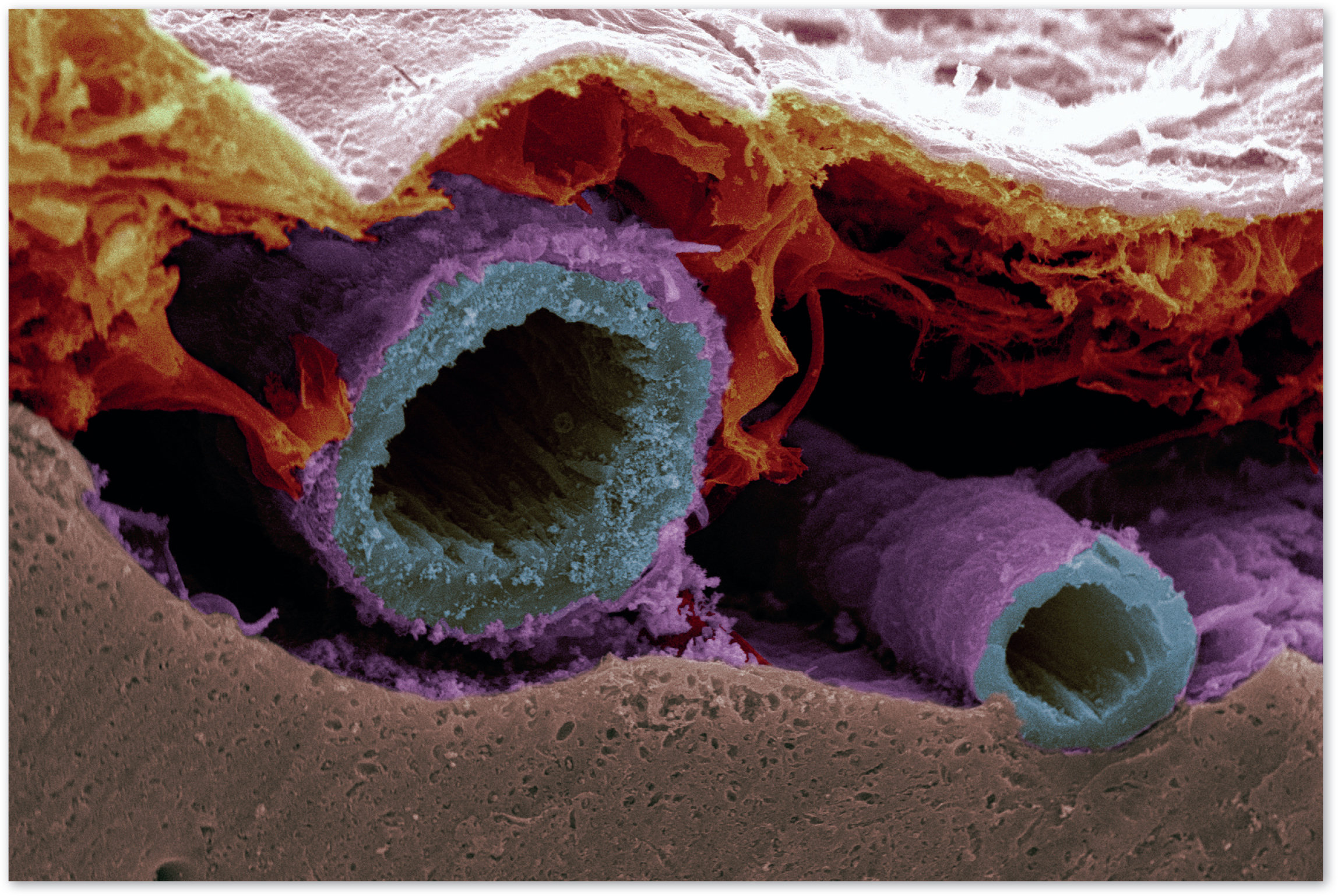
An image of a normal artery at the surface of a rat's brain was taken with a powerful scanning electron microscope. These ‘subarachnoid vessels’ supply blood to the brain and also act like a drain to remove toxic waste products. The runner-up winner of the 2018 British Heart Foundation's annual image competition, Reflections of Research, Matt MacGregor Sharp, and his team are trying to show that failure to remove waste by these vessels is one of the underlying causes of vascular dementia.
The researchers took the image using a technique called ‘freeze fracture’, where tissue or cell samples are frozen and then split apart to reveal the hidden layers within the sample so they can be studied in extreme detail. Sitting above the brown brain tissue, the artery appears blue, and its surrounding layer, the pia mater, is shown in purple.