

This 64-year-old man had an anterior ST elevation myocardial infarction (STEMI) during the first COVID-19 lockdown. He had an episode of ventricular fibrillation (VF) during his primary percutaneous coronary intervention (PCI) procedure and was promptly defibrillated into sinus rhythm. He has a single stent inserted in his mid-left anterior descending coronary artery. His cardiac rhythm remained stable thereafter. His echocardiogram showed moderate left ventricular systolic dysfunction.
This man's post myocardial infarction (MI) cardiac rehabilitation follow-up was suboptimal due to the impact of the pandemic on traditional face-to-face clinics.
He was subsequently re-admitted with central chest pain on three separate occasions over the next 9 months. On each occasion, his 12-lead ECG was unchanged and his high sensitivity troponin assay was negative.
He has now presented to his GP with ‘palpitations’. He has a feeling that his heart has momentarily stopped and, on other occasions, is aware of his heart thumping in his chest.
His vital signs were as follows:
- Blood pressure: 158/92 mmHg
- Pulse: 90 beats per minute with occasional irregularity
- Respiratory rate: 16 breaths per minute
- He was apyrexial
- SpO2 on air: 97%.
His GP recorded a 12-lead ECG and it was unchanged from the most recent recordings.
His GP suspected ectopic activity and arranged for a short period of cardiac monitoring at home. The ECG rhythm strip in Figure 1 was recorded while he was experiencing symptoms.
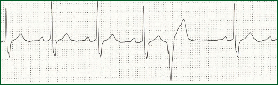
Interpretation of the ECG rhythm
- The heart rhythm is regular
- The heart rate is approximately 65 beats per minute
- The rhythm is sinus rhythm
- The 5th beat in this sequence is a ventricular ectopic.
Ectopic beats or extrasystoles are extra beats that come early in the cardiac cycle. They were first described by the Chinese physician, Pien Ts'lo, in 600 BC. Ectopic activity is detected in 40–75% of healthy people on 24-to 48-hour monitoring.
Ventricular ectopics or premature ventricular contractions (PVCs) are identified as premature beats with a wide and bizarre QRS morphology. They usually have discordant ST segment and T wave, i.e the QRS and ST segment and T wave are deflected in opposite directions.
As in this case, many patients describe a sensation of their heart momentarily stopping. This is because they are aware of the compensatory pause that occurs with ventricular ectopics. In the example above, measure the distance between the beat before and the beat after the ectopic. This will be the same as two normal cardiac cycles.
In normal hearts, ventricular ectopics are usually of no clinical significance. In most cases, patients are largely asymptomatic. This man was reassured and enrolled onto a robust cardiac rehabilitation and group exercise programme. His symptoms have largely settled since.