Immersion in cold water is a common event. Drowning, which is defined as death caused by an impairment of respiratory function from immersion in liquid, is a major cause of death worldwide (World Health Organization, 2003). In most countries, it reflects the third most common cause of unintentional death in adults and the second most common in children, with approximately 500 000 immersion-related deaths occurring internationally every year (Bierens et al, 2002; Shattock and Tipton, 2012). In the UK there were 420 water-related deaths in 2010 with the highest number (52%) occurring in inland waters such as rivers, canals, lochs and lakes (National Water Safety Forum, 2012). With seawater temperature varying between 15–18ºC at its warmest, the threat of sudden cold water immersion in the UK is a real one. Even for those patients who survive to the Emergency Department (ED), half will require hospitalisation or transfer to further care, with non-fatal injuries resulting in neurological deficits including a permanent vegetative state (Cummings and Quan, 1999). In previous years most immersion victims were treated as drowning cases and managed appropriately. However, with the increased use of items such as the lifejacket, immersion hypothermia has become a common consideration too (Golden and Rivers, 1975). Despite this, and accidental hypothermia being associated with significant mortality and morbidity, the exact incidence of immersion hypothermia remains unknown (Brown et al, 2012). In fact, cold water immersion more generally is still thought to be poorly understood by the general public and even by many outdoor experts (Giesbrecht and Wilkerson, 2006).
An immersed patient is characterised by Tilton (2010) as someone whose airway remains above the surface allowing them to breathe, and is to be differentiated from submersion, where the patient is unable to breathe for an extended amount of time due to being below the surface of the water. Stereotypically, such an event might occur following a fall through ice or falling from a boat, but coinciding trauma, drug and alcohol intake, and suicide intent is also to be considered in each case (Fleisher and Ludwig, 2010). Nonetheless, all instances are severely complicated in the presence of colder water. Among academic texts, cold water is generally considered to be below 15ºC, with the World Health Organization (2003) recognising that exposure to water below this temperature can result in a ‘debilitating shock response’, as well as the co-existence of hypothermia and drowning. Whilst four physiological stages of cold water immersion were known, it was Golden and Hervey in 1981 who presented these in more detail, offering a greater explanation into the causes of death in these patients.
The four stages
Golden and Hervey's four stage theory (Golden and Hervey, 1981) begins during immediate contact with the water. On initial immersion, an autonomic reflex is triggered involving uncontrollable hyperventilation, tachycardia and hypertension. This first stage is thought to last up to 5 minutes before the body's response changes to one of rapid muscle and nerve cooling, known in this context as ‘cold incapacitation’. During the 5–30 minutes of stage two, muscle activity becomes so ineffective that without the use of a lifejacket or similar buoyancy aid, the patient loses the ability to stay afloat. It is at this point (stage three) that hypothermia becomes life-threatening, with death occurring sometime after 30 minutes of immersion in icy water. If death is averted, the final stage happens in the presence of rescue. Known as circum-rescue collapse, during extrication the immersed patient is at risk of cardiac arrest—in part due to changes in blood pressure coupled with the potential for a ‘cold heart’. The detrimental effect of these stages is obvious, with over half of drowning incidents occurring in stages one and two (Brooks et al, 2008).
Physiology
In order to understand Golden and Hervey's four stages in greater detail it is important to realise the body's normal physiology, especially in respect of the systems that are most affected during immersion—namely, the respiratory, nervous and cardiovascular systems. Of these, it is the respiratory system that perhaps has the most obvious effect. Comprising of the upper and lower respiratory tract and aided by intra-thoracic pressure changes caused by the muscles of respiration, its primary purpose is to facilitate gas exchange while also having some metabolic responsibilities (Ward et al, 2010). In normal respiration, central and peripheral chemoreceptors recognise a reduction in blood pH. This results in nerve impulses from the respiratory centre stimulating the diaphragm and intercostal muscles via the intercostal and phrenic nerves. Consequently, air moves down the concentration gradient allowing ventilation and oxygenation to happen (Robinson and Scullion, 2009). In the submersed patient, a severe interruption of this process occurs. Wet drowning involves inhalation of fluid into the lungs causing pulmonary vasoconstriction, pulmonary hypertension, decreased lung compliance, surfactant washout and subsequent atelectasis. As gas exchange is halted by the process of drowning, hypoxia, hypercapnia and respiratory acidosis develop (Wyatt et al, 2012). Those patients who suffer near drowning episodes also run the risk of secondary drowning: a deterioration of a rescued patient in which electrolyte changes facilitate fluid shift across the alveolar membrane. This form of non-cardiogenic pulmonary oedema can occur up to 72 hours after the near-drowning episode, and paramedics should be vigilant for worsening in a near drowned patient (Adelman, 2011). In terms of application, not all drowning victims will present with pulmonary oedema or evidence of significant aspiration. Dry drowning is thought to be attributed to 10–20% of deaths, with persistent laryngospasm resulting in asphyxia and an ‘immediate outpouring of mucus, froth and foam’ from the mouth (Wyatt at al, 2012), something that should be considered during airway assessment and management of an immersed patient.
In addition to respiratory complications, life-threatening consequences can develop as a result of neurological changes. The autonomic nervous system plays a fundamental role in the maintenance of homeostasis and self-protection (Timby, 2009). However, it is this very subdivision that is thought to have a harmful impact upon the patient at the point of initial immersion, termed the ‘cold shock response’.
As the patient is immerged, the sympathetic branch initiates a systemic response involving the cardiovascular and respiratory systems. Peripheral cold receptors stimulate the respiratory centre via thermoafferent nerves (Goode et al, 1975) and uncontrollable hyperventilation occurs with the patient gasping, unable to hold their breath. Aspiration and/or drowning may occur at this point depending on a number of factors including water temperature and body movement (Tipton, 1993). Hyperventilation, reaching nearly five times that of normal values, results in a reduction of carbon dioxide partial pressures and causes a relative respiratory alkalosis. This in turn leads to cerebral vasoconstriction with subsequent effects on mental status including a loss of consciousness (Wittmers and Savage, 2002).
Similarly, the sympathetic cardiovascular effects of immersion are equally concerning. The sudden exposure to cold water stimulates the secretion of adrenaline, noreadrenaline and cortisol, increasing stroke volume, heart rate and peripheral resistance. Profound vasoconstriction combined with an increased cardiac output mean a normotensive individual becomes immediately hypertensive, with a reported systolic increase of 45 mmHg within 1 minute (Bove, 2004). Hydrostatic pressure exerted on the body during immersion can further increase blood pressure and the shunting of blood to the core (Datta and Tipton, 2006). For these reasons it is suggested that the ‘cold shock response’ is perhaps the most deadly stage of cold water immersion. Those patients with underlying heart disease, hypertension or other cardiovascular risk factors are at an increased chance of heart attack or stroke, with even young people in the presence of angiographically normal coronary arteries being known to suffer acute myocardial infarctions (Raguz et al, 2008).
Autonomic conflict
Certainly, with regard to the neurological effects on the cardiovascular system, the most interesting recent development is the proposal of an ‘autonomic conflict’ theory. Whilst it is well recognised that sudden cold water immersion may induce immediate cardiac arrhythmias (Olsen et al, 1962; Schmid et al, 2009), it is Shattock and Tipton (2012) who suggest that a powerful clash may exist between the sympathetic cold shock response and that of the parasympathetic mammalian diving reflex. The diving response, seen in all marine and land vertebrates, appears to exist to conserve oxygen during prolonged time underwater. Instant cooling of the cold receptors in the face innervates the maxillary and ophthalmic branches of the trigeminal cranial nerve. This excites the cardiac vagal nerve whilst inhibiting central respiratory neurones and stimulating vasoconstrictor nerves. The result is a significant bradycardia associated with bradypnoea and vasoconstriction at the point of initial immersion (Angell-James and Daly, 1975) —an effect that opposes the sympathetic cold shock response. Shattock and Tipton (2012) go on to explain that if varying degrees of parasympathetic vagal stimulation occur in the presence of underlying sympathetic innervation, then potentially life-threatening arrhythmias can result. Indeed, in those patients where this dual activation occurs, 62–82% develop arrhythmias which typically include paroxysmal ventricular tachycardia, premature atrial complexes, atrio-ventricular block and sinus bradycardia (Tipton et al, 1994; Datta and Tipton, 2006). Given such profound effects, paramedics should be mindful of arrhythmias and understand the importance of thorough and continual cardiac monitoring.
Whilst Shattock and Tipton's theory is certainly one of immense interest, in the pre-hospital care setting it serves less relevance due its instant nature. In most cases paramedic-led assessment would not take place until long after initial immersion, especially considering the sometimes remote areas in which immersion might occur. Even with a timely ambulance response, such immediate incapacitation caused by sudden cardiac arrhythmias would likely lead to agonal respiration, inhalation of water and death from drowning (McDonough at al, 1987), all of which would take priority during the ‘single most important steps’ of re-establishing ventilation and relieving hypoxia (Boffard et al, 2001). Therefore, it is perhaps Golden and Hervey's later stages of immersion that are more relevant at the point of initial assessment, due mostly to their insidious onset.
Assessment
In terms of assessment, paramedics would be more likely to encounter symptoms of the ‘cold incapacitation’ or hypothermic stages of immersion, because of their delayed effects. During the second stage, which typically lasts from 5–30 minutes, vasoconstriction of the peripheries pushes blood centrally in an attempt to maintain core body temperature. This has the effect of leaving exposed limb tissues with only a minimal supply of warm blood, allowing a rapid cooling of nerve and muscle tissue (Giesbrecht and Wilkerson, 2006). How quickly this happens evidently depends on the temperature of the water, with action potentials slowing on average 15 m/s for every 10ºC drop in local temperature (Whyte et al, 2005). Even before hypothermia becomes problematic, dexterity, speed of movement and strength are all significantly reduced until the patient becomes increasingly exhausted. In a review into current literature, Ducharme and Lounsbury (2007) reflect this by concluding that swimming failure develops before systemic hypothermia, allowing victims to swim 800–1 500m in water of 10–14ºC before becoming incapacitated by cold. In respect of assessment at this stage, such a negative impact upon neuromuscular activity is likely to result in abnormalities during a neurological examination, especially with regard to the peripheral nervous system. Reduced sensation, slow movement, dysarthria, slow tendon reflexes, and/or mal-coordination might be expected, making the differentiation between this, hypothermia and other differential diagnoses (including head injury) difficult, owing to their similar presentations and the ability of one masking the other (Wittmers and Savage, 2002; Søreide and Grande, 2005).
Hypothermia and thermoregulation
While cold incapacitation victims may bring about challenges for the paramedic, it is hypothermia that mostly dominates considerations during assessment. Immersion hypothermia, which makes up one of three main types of accidental hypothermia, results in heat production being overwhelmed by sudden cold stress (Moulton and Yates, 2012). Water causes heat loss 25 times faster than exposure to air at the same temperature (Sanders et al, 2012) and loss of core body temperature may arise within 30 minutes (Tilton, 2010). In water of 10ºC, an average clothed individual will have a steady decline in core temperature and die from hypothermia or drowning within 4 hours (Golden and Tipton, 2002). To prevent this detrimental change, the body is well known for attempting to minimise heat loss. Thermoregulation in a healthy person is normally effective, maintaining a core body temperature of 37.2–37.6ºC, even in dry air temperatures as low as 13ºC (Rutty, 2012). Central control of temperature is regulated by the anterior hypothalamus, which sits inferior to the thalamus, capping the brain stem. Here, with a drop of temperature, heat sensitive neurons release thyrotropin hormone causing the pituitary gland to secrete thyroid stimulating hormone. This in turn causes the release of thyroxine, increasing cellular thermogenesis. In addition, nervous innervation produces piloerection, shivering and vasoconstriction; all in an attempt to retain heat for normal cell function (Sherwood, 2008).
If a normal temperature is not maintained then symptoms develop which can indicate the severity of hypothermia. Understanding this is fundamental to pre-hospital assessment and management as peripheral temperature measurement is inaccurate and is made even more difficult due to standard thermometers not detailing temperatures of below 34.4ºC (Partridge et al, 2012). During the earliest stage of hypothermia, thermoreceptors found in the subcutaneous tissue and skin promote localised vasoconstriction. The hypothalamus is then stimulated to produce thyroid-stimulating hormone and adrenocorticotropic hormone, which act upon the thyroid and adrenal glands respectively. In addition, the hypothalamus promotes shivering, which generates heat from skeletal muscle, ultimately caused by the breakdown of high-energy phosphate bonds. Prolonged vasoconstriction may result in acidosis, reducing the efficacy of catecholamine production—something that increases inversely with water temperature (Schmid et al, 2009). Clinically, mildly hypothermic patients (32–35ºC) present shivering with tachycardia and fatigue. Early stimulation of the respiratory centre also results in tachypnoea (Partridge et al, 2012; BMJ Evidence Centre, 2012a). As hypothermia progresses, continual ECG monitoring will show the development of atropine-resistant bradycardia and the potential for a J wave, both as a result of depolarisation and repolarisation abnormalities. Respiratory rate and tidal volume decrease, leading to an increase in anatomical dead space and pulmonary oedema (Mallet, 2002). At this stage of ‘moderate hypothermia’ (28–32ºC) the body's energy production decreases and the compensatory shivering mechanism stops, with patients presenting with a decreased level of consciousness (Partridge et al, 2012). Finally, severe hypothermia (below 28ºC) results in an unresponsive patient with a fatal appearance. The patient may display arrhythmias and present in cardiac or respiratory arrest. Owing to the neuroprotective qualities of hypothermia and its ability to suppress the pathways leading to cell death, successful resuscitation from this state is well known, with one case report documenting the recovery of a patient from a core temperature of 13.7ºC (Gilbert et al, 2000; UK Resuscitation Council, 2010). Resuscitation of the immersed patient should therefore not be dismissed without careful consideration, even if physical assessment reveals apparent futility. Current ambulance resuscitation guidelines reflect this, advocating advanced life support in patients who have been submerged for up to 1.5 hours (Joint Royal Colleges Ambulance Liaison Committee (JRCALC), 2013).
Immediate assessment and management
In reality, the pre-hospital assessment of any immersion victim is likely to be challenging from the outset. Adopting a structured approach is vital, with scene safety remaining the highest priority. In fact, the very nature of the incident suggests a potentially hazardous scene, making it essential that a ‘reasonable degree’ of safety is sought during immersion incidents (American Academy of Orthopaedic Surgeons, 2010a). Beyond this, a standardised ABCDE approach to the assessment and management of the immersion victim is difficult due to the possibility of the patient remaining immersed. While many sources advocate ‘immediate intervention’ and ‘early, rapid intubation’ in near drowned patients (Mason, 2001; BMJ Evidence Centre, 2012b), little consideration is given to the harmful effects of the fourth stage of cold water immersion, circum-rescue collapse. Indeed, if the patient remains immersed, assessment and management priorities may be altered as hasty actions at this point may cause a rapid deterioration, attributing to 17% of immersion deaths (Tochihara and Ohnaka, 2005). It is this physiological stage of cold water immersion that has to be the most relevant in terms of immediate patient management. Sudden removal from the water is known to initiate instantaneous hypotension, owing in part to the abrupt reduction of hydrostatic squeeze that the water created during immersion. The body's ability to compensate for this drop in blood pressure is likely to be reduced in the presence of hypothermia. A cold myocardium may struggle to increase cardiac output whilst an increase in peripheral resistance may be ineffective due to already profound vasoconstriction (Steinman and Giesbrecht, 2001). Furthermore, mental relaxation, decreased levels of stress hormone secretion and vasodilation at the point of extrication may also contribute to deterioration during circum-rescue collapse (Golden and Hervey, 1981). Such a sudden decrease in venous return to the heart may precipitate ventricular fibrillation and myocardial ischaemia due to a decrease in coronary artery perfusion (Golden et al, 1997). It is for this reason that patients should be removed from the water gently with extreme caution, preferably horizontally, while minimising the patient's physical activity (Steinman and Giesbrecht, 2001).
Given this information, it seems sensible that a high degree of thought should surround the ABC assessment of the immersed patient. It should perhaps be the outcome of the primary survey that dictates patient management if they remain immersed. For example, if the patient presents with an airway occlusion or in respiratory or cardiac arrest, then rapid extrication from the water would be appropriate. In contrast, sudden, vertical removal of the conscious patient may lead to a rapid, fatal decline (Harries, 2003).
Further assessment and management
When arriving at the patient, a rapid and methodical primary survey including consideration of spinal immobilisation and mechanism of injury is something advocated in all major life support texts, regardless of current immersion or not (McQuillan et al, 2008). The airway should be assessed for fluids, bleeding and signs of obstruction including gurgling, stridor, accessory muscle use, tracheal tug and a reduced level of consciousness (Bhangu et al, 2010). Drowned or near drowned patients can be expected to vomit (Tilton, 2010), with comatose patients requiring immediate intubation and oxygenation (BMJ Evidence Centre, 2012b). Initial high concentrations of oxygen are required for hypothermic and near-drowned patients (O'Driscoll et al, 2008) followed by assessment of ventilatory status. Thorough auscultation is key and may reveal a number of abnormalities including pulmonary oedema and/or diminished breath sounds (American Association of Orthopaedic Surgeons, 2010b). Paramedics should be encouraged to use capnography where possible and not rely on pulse oximetry as it can vary significantly in the immersed patient (Kolb et al, 1999). Ventilation via a bag valve mask may be necessary to relieve hypoxaemia and is thought to be the single most important intervention for a drowned or near drowned patient (Soar et al, 2010).
Assessment of circulation can then be carried out. In the hypothermic patient this might be difficult as heart rate (and respiratory rate) may be exceptionally slow, with the potential for absent peripheral and central pulses (Mahadevan and Garmel, 2005). Management at this point is controversial. Guidelines are clear that, in the presence of an unresponsive patient with agonal or absent breaths, CPR should be commenced (UK Resuscitation Council, 2010). However, other texts warn against CPR in hypothermic patients if electrical complexes are present on the ECG, irrespective of a palpable pulse. This is due to the thought that cardiac output probably still exists and that the trauma of chest compressions is likely to precipitate ventricular fibrillation of the already irritable cold heart (Goodenberg, 2000; Schwartz et al, 2008). While the American Heart Association and The European Resuscitation Council recognise the difficulties in assessing circulation, there appears to be little evidence to support withholding CPR in the absence of a central pulse. Both guidelines advocate starting CPR if there are any doubts about the presence of a pulse and recommend looking for signs of life for up to 1 minute before determining there is insufficient cardiac output (Soar et al, 2010; Vanden Hoek et al, 2010). Once the decision has been taken to attempt resuscitation in the severely hypothermic patient, non-essential drugs should be avoided because of an unpredictable metabolism leading to toxicity once the patient is rewarmed (Peitzman et al, 2008). In the presence of severe hypothermia, the latest JRCALC guidelines suggest limiting defibrillation attempts to three and withholding intravenous drugs until body temperature reaches above 30ºC. The dose interval should then be doubled until a temperature of 35ºC is achieved (JRCALC, 2013).
Regardless of the patient's presentation, the underlying management principles of the cold patient remain the same: prevention of core temperature decline, moderate core rewarming and transport to definitive care (Steinman and Giesbecht, 2001). A careful search for other disorders during a secondary survey is also important, including looking for diabetes, sepsis, intoxication and physical injures (American College of Surgeons, 2008). Cold injuries including frostbite may be present but should not take precedence over the management of hypothermia (Wyatt et al, 2012). Preventing heat loss by removing wet clothing and moving the patient to a warm environment is fundamental, as is minimising heat loss during exposure and examination. Regular reassessment to detect temperature change is also essential (Long et al, 2005). Despite such obvious, common sense actions, pre-hospital treatment of hypothermia lacks a strong evidence base, with only one or two pre-hospital guidelines detailing specific patient management. In the UK, the JRCALC (2013) offers only minimal advice on rewarming patients out of hospital, with certainly no mention of the differing methods.
Rewarming hypothermic patients
Hamilton and Paton (1996) recorded seven different ways in which mountain rescue teams rewarm patients, with chemical pads, spontaneous rewarming and hot water bottles being among the most common. These can be divided into three groups: external passive warming (e.g. removal to a warm environment), external active warming (where heat is transferred directly to the skin), and active core rewarming (involving heat being passed to the body's core) (Schwartz et al, 2008). It is the more recent, latter group which shows most potential for the future treatment of hypothermia, with extracorporeal rewarming being demonstrated as the most effective method, increasing core temperature 1–2ºC every three to five minutes (Mccullough and Arora, 2004). However, the practical application of this in the pre-hospital setting is inappropriate, owing to the common problems that can occur during restoration of normal temperature. In a retrospective cohort of 84 patients suffering moderate to severe hypothermia, 79 developed pulmonary, renal or neurological complications during rewarming (van der Ploeg et al, 2010). Steinman and Geisbrecht (2001) recognise this stating that, although extracorporeal rewarming might be a management tool of the future, aggressive rewarming in the field is currently contraindicated due to the inability to diagnose or manage the potential complications. Indeed, care should be taken as too rapid a rewarming may bring about vasodilation, cerebral or pulmonary oedema and rewarming acidosis (Mulcahy and Watts, 2009; Wyatt et al, 2012).
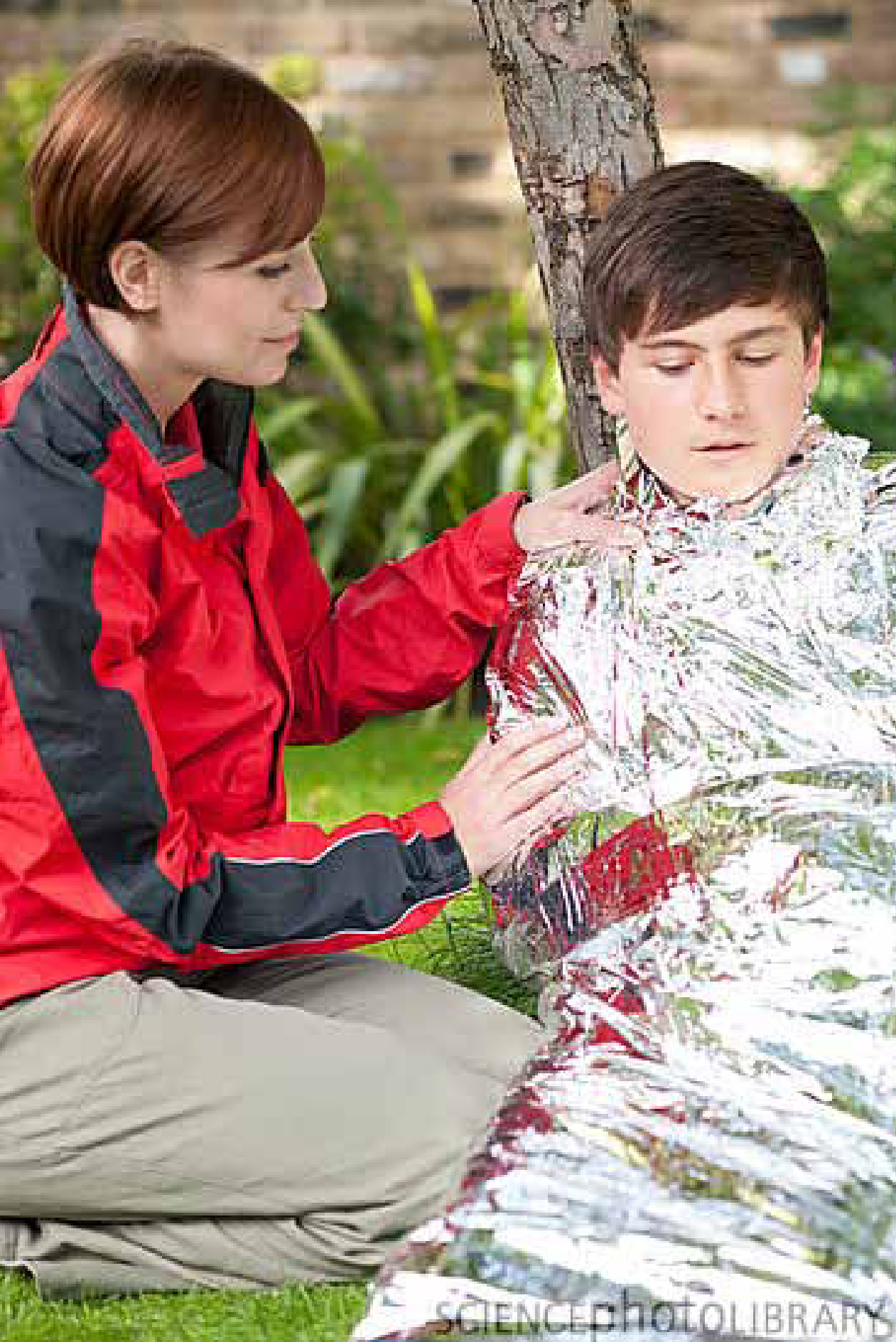
In UK paramedic practice, the choice of rewarming method is simplified due to a lack of availability of anything other than foil and cotton blankets (Figure 1), a warm ambulance environment and warmed intravenous fluid. Certainly in mild and moderate hypothermia the passive influence of a warm ambient temperature and multiple layers of blankets is appropriate, evidence-based management (American Heart Association, 2005). However, evidence for warmed fluid is less clear and the minimal human data that does exist surprisingly shows no real benefit. In a study into the efficacy of warming devices in 174 trauma patients, warmed intravenous fluid administration was shown to produce a 0.2ºC drop in body temperature (Watts et al, 1999). Participants were subject to a range of interventions, with only chemical hot packs demonstrating an increase in temperature. Some explanation might be sought due to the titration of the fluid to maintain a systolic blood pressure of 90 mmHG, leading to variable amounts of fluid and infusion rates. Furthermore, tympanic measurement (the method used for this study) is undependable and has been shown to be clinically problematic, with discrepancies of 0.3ºC (Morgan et al, 2007). Nonetheless, whilst studies on dogs have demonstrated warm IV fluid to be effective at warming hypothermic patients (Sheaff et al, 1996), The European Resuscitation Council stress that heated IV fluids are inefficient, raising core body temperature by 0.3ºC for every one litre of infused 40ºC fluid (Paal et al, 2006). Of significance, they go on to recommend that transport to definitive care should not be delayed due to attempts at core rewarming (Soar et al, 2010). Management of the hypothermic patient should therefore hold a less invasive approach, with warmed IV fluid being reserved only for the hypotensive effects of cold water immersion.
Conclusions
Understanding the four physiological stages of cold water immersion is fundamental to informing good clinical judgement. The paramedic must be particularly aware of the cold incapacitation, hypothermic and circum-rescue collapse phases, as these may be encountered in the field, directly affecting patient presentation and treatment. Hypothermia and drowning often go hand in hand and can present challenges to emergency medical services. Assessment and treatment should focus around a structured ABCDE approach, with good ventilation and oxygenation remaining a cornerstone of immersion management. A secondary survey must not be overlooked because co-existing injuries and illness can also be present. Hypothermic patients' priorities lie in moderate core rewarming and transport to hospital. A warm environment with layers of blankets should be provided with regular reassessment of temperature. Heated intravenous fluids are not effective as a rewarming method, but play a key role in the management of the hypotensive rescued patient. The neuroprotective qualities of hypothermia must not be underestimated and a low threshold maintained for initiating basic and advanced life support.