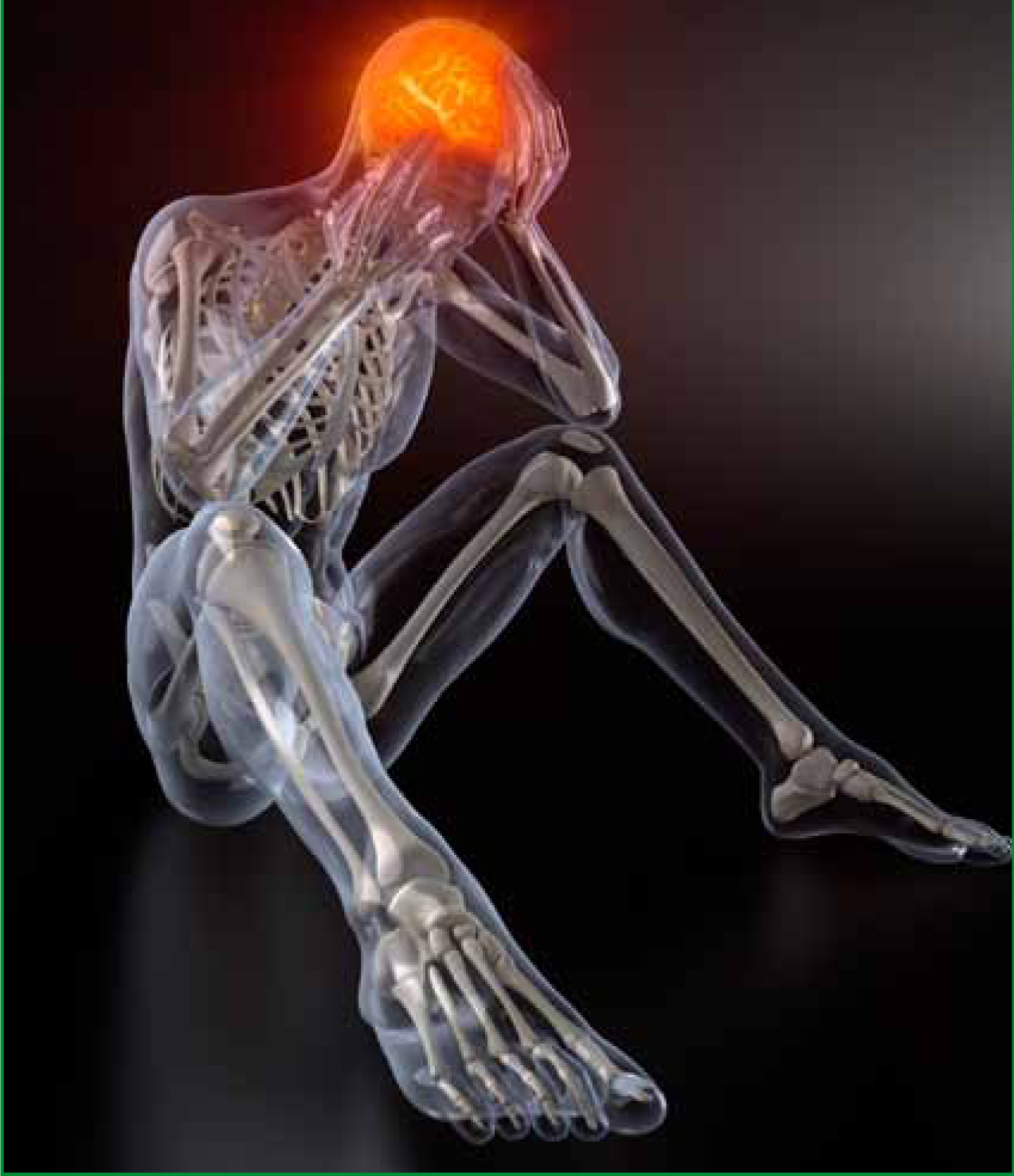
An emergency call was received by Ambulance Control in response to an adult female following a suspected, although unwitnessed fall from a horse. The rider was found by fellow equestrians crawling around a field in an agitated and confused state, with little available information regarding the events proceeding the suspected fall. From the limited history available, it appeared more likely than not, that the patient had suffered a cerebral insult either prior to, or during a fall from her horse. On arrival of the pre-hospital helicopter emergency medical services (HEMS) team consisting of two critical care paramedics (CCP) and a HEMS doctor; the patient was found agitated, combative, mumbling incoherently and unable to comprehend simple commands from the medical team.
Patient assessment and clinical findings
Immediate manual inline immobilisation was initiated to prevent further secondary injuries from a possible undiagnosed c-spine fracture, while a primary survey was undertaken. This survey provides a rapid basic clinical assessment to ascertain immediate threats to life, but proved extremely difficult to undertake due to the agitated behaviour displayed by the patient. The primary survey revealed that there was no catastrophic haemorrhage; the patient maintained a patent airway; was tachypnoeic, and appeared well perfused; with a palpable radial pulse. Although, a significantly impaired level of consciousness was observed, which immediately raised suspicions of a potential moderate to severe traumatic brain injury (TBI). However, with no significant damage to the rider’s helmet and in the absence of any visible external injuries to the face, head or scalp to indicate a TBI, the extent of agitation at this stage appeared inconsistent. This inconsistency prompted the medical team to consider other potential diagnoses for the patient’s behaviour, such as a primary cerebral event associated with pre-existing cerebral pathology, hypoxic brain injury, hypoglycaemia, alcohol consumption and/ or drug abuse.
Although, a TBI with a space occupying lesion, such as an intracranial bleed or contusional injuries, initiating a rise in intracranial pressure, and the marked pupillary changes observed, could not be excluded. The need to remain astute to other, less-common conditions was paramount, and not to consider TBI as the sole diagnosis.
No other obvious major time-critical chest, abdominal or pelvic injuries were identified at that stage during a gross visual examination, and the patient was moving all four limbs with no obvious long bone fractures. Gathering further information relating to pertinent past medical and pharmaceutical history proved impossible from either the patient or bystanders at the scene.

A more detailed secondary examination proved extremely challenging and difficult to undertake without further clinical invention to aid the management of this patient. Therefore, sedation of the patient was undertaken to facilitate this.
A more comprehensive assessment revealed contusions to the anterior chest wall and unilateral differences in pupil size (left=6 mm/right =2 mm) which were suggestive of time-critical neurological pathology.
Capacity and consent
An integral part of the initial management plan was to gain informed consent to examine and treat the patient. It is essential when undertaking clinical assessments or providing treatment to any patient, that their consent to any medical procedures is obtained, therefore the medical team had to consider whether the patient had sufficient capacity in this case. The Mental Capacity Act (UK Parliament, 2005) states that ‘a person must be assumed to have capacity unless it can be established that they lack capacity’, and that ‘everyone with capacity has the right to give or withhold their consent to treatment’. The patient’s agitated behaviour was suggestive of a lack of capacity, due to the inability to assimilate and retain the information being provided by the medical team, or rationalise the consequences or potential implications for non-compliance to instructions provided. The lack of capacity was evidenced by the presence of confusion, agitation, and alterations in neurological status, which are all clinical features commonly associated with significant primary or secondary traumatic brain injuries, or intracranial cerebral events. The Mental Capacity Act (UK Parliament, 2005) was also applied in this case. This is applicable when:
‘…a person lacks capacity in relation to a matter, if at the material time they are unable to make a decision for themselves because of an impairment of, or a disturbance in the functioning of, the mind or brain.’ (Mental Capacity Act, 2005)
Assessment of the patient’s neurological function, including cognitive behaviour and motor function was achieved by the application of the Glasgow Coma Scale (GCS), a neurological scoring system ranging from 3 (unconscious) to 15 (alert and orientated), which provides an objective test for measuring the conscious state of a patient. Following application of the scoring system by the medical team, the patient’s GCS was calculated at 9/15, specifically: Eyes (E)=3, Motor (M)=4 and Verbal (V)=2. Based on this finding it was deemed that the patient lacked capacity under the Mental Capacity Act (UK Parliament, 2005) to consent to or refuse treatment. It was therefore incumbent upon the medical team to act out of necessity to provide potentially lifesaving interventions in order to safeguard the best interests of the patient, in accordance with the Mental Capacity Act (UK Parliament, 2005). The Joint Royal College Ambulance Liaison Committee ( JRCALC, 2006) states that ‘in adults who usually have capacity may, especially in emergency situations, become temporarily incapable’ and ‘it is permitted to apply treatments that are necessary and no more than is reasonably required in the patients best interests pending recovery of capacity’, which ‘includes any action taken to preserve the life, health or well-being of the patient’.
The use of physical restraint
After several unsuccessful attempts at verbal communication and reasoning, moderate physical restraint was deemed necessary to administer treatment. The decision to restrain patients is often complicated and sometimes anxiety provoking for all concerned, especially in time-critical emergency situations were insufficient time is available to undertake a full consultation with a patient who is reasonably believed to lack capacity. Therefore, having established that the patient lacked capacity, reasonable and proportionate force was applied to prevent further self-.harm and initiate treatment, in accordance with the Mental Capacity Act (UK Parliament, 2005).
This form of restraint proved distressing for friends and bystanders who were witnessing the incident, but potentially extremely unpleasant for the patient too. Verbal reassurance continued during the restraint, in attempts to partially alleviate any distress or associated anxieties, but also in support of the patient’s physical and emotional wellbeing during the intervention. However, the Association of Anaesthetists of Great Britain and Ireland (AAGBI) Pre-hospital Anaesthesia Guidelines (2009) do not advocate the use of restraint in such cases, due to the potential exacerbation of the patient’s clinical condition, commonly associated with ‘an increase in blood pressure, which may increase intracranial pressure or promote non-compressible bleeding’ in traumatic brain injured patients. The AAGBI also state that restraint may ‘cause further injuries or exacerbate spinal injuries’, and makes ‘reliable pre-oxygenation difficult’ in patients requiring RSI or procedural sedation. Although, they do acknowledge that ‘every case should be assessed and managed on its own merits’ and that the pre-hospital environment is often ‘challenging’. While acknowledging the validity and value of such points, immediate clinical management of the casualty was essential to prevent further self-harm, and was achieved using minimal force as necessary to initiate emergency treatment, in accordance with the Mental Capacity Act (UK Parliament, 2005).
Sedation
The use of reasonable and proportionate force enabled the medical team to gain prompt intravenous (IV) access using a large bore cannula inserted in the left antecubital fossa (ACF), and administer small aliquots of midazolam intravenously, in accordance with The Air Ambulance Service (TAAS) Clinical Standard Operating Procedure (CSOP) (CSOP, 2010) in order to sedate the patient sufficiently to facilitate effective management. Midazolam was administered cautiously and titrated to effect in order to prevent the initiation of a secondary brain injury due to an iatrogenic initiation of systemic hypotension and fall in mean arterial pressure (MAP), resulting in poor cerebral perfusion pressures (CPP).
Therapeutic doses of midazolam appeared to work sub-optimally, and while rendering the patient ‘more manageable’, additional doses may well have precipitated hypotension and airway compromise. Therefore, the decision to supplement any further doses of midazolam with ketamine was taken. Ketamine was chosen due to its rapid onset of action, and the anticipated progression towards undertaking a formal RSI using ketamine as the induction agent.
Patient monitoring was immediately applied to ensure patient safety following sedation, in accordance with the AAGBI pre-hospital Anaesthesia Guidelines (AAGBI, 2009) and our own organisational TAAS CSOP 013. For comparison, the initial clinical observations post-sedation are documented in Table 1. Oxygen was applied simultaneously to the monitoring and 15 litres/min was initially administered via a close fitting non re-breathable oxygen mask with reservoir bag and oxygen saturations maintained between 94–98 %, as advocated in the British Thoracic Society Guidelines (O’Driscoll et al, 2008) adopted by the Joint Royal Colleges Ambulance Liaison Committee ( JRCALC) in April 2009. These guidelines consider the optimal saturation range to be 94–98 % to reduce the potential adverse affects of hyperoxaemia, which is closely correlated to vasoconstriction in the capillary beds, frequently resulting in reduced blood flow and poor perfusion in damaged tissues and end organs. Once stabilised, the patient was then placed in a LESS® Thermal Blanket— this is a thermal bubble wrap device used to preserve body temperature by minimising heat loss. Hypothermia is commonly associated with coagulopathic complications and poor outcome in poly-trauma patients, but seen to have beneficial effects in isolated head injuries, however, prevention of heat loss in all anaesthetised patients is paramount, as anaesthetised patients lose the ability to physiologically regulate their own body temperature through shivering.
‘The prevention of hypoxia and hypercarbia is paramount, and are considered as main contributory factors to secondary brain injuries. ’
Clinical management and rapid sequence intubation
The early decision to progress to a formal pre-hospital Rapid Sequence Intubation (RSI) was based largely on providing ventilatory support, optimising oxygenation, maintaining an adequate MAP to reduce the likelihood of secondary hypoxic brain insults resulting from poor or inadequate cerebral perfusion. This intervention involves combining an appropriate induction, paralytic and maintenance agent to render a patient rapidly unconscious and flaccid in order to facilitate endotracheal intubation, therefore minimising the risk of aspiration and to facilitate optimal ventilatory support. The pharmacology used to facilitate RSI in this case, was ketamine as an induction agent, suxamethonium and rocuronium as paralytic agents, and morphine and midazolam as maintenance agents, all of which were deemed appropriate pharmacology by the HEMS team, in accordance with the TAAS CSOP013, the Brain Trauma Foundation, and the AAGBI Guidelines.
The patient’s clinical presentation was indicative of the need for urgent diagnostic CT imaging, ideally within one hour of arrival in the emergency department (ED). Therefore, initiating anaesthesia in the pre-hospital arena would vastly speed up the transition from hospital arrival to diagnostic imaging and early neurosurgical intervention, optimising the clinical care this patient received, which is advocated in the National Institute for Health and Clinical Excellence (NICE) Head Injury Management Guidelines (2007). Laryngoscopy revealed a post-commissural grade two view, through which a size 7.0 endotracheal tube was successfully placed in the trachea. Chest auscultation and end-tidal (EtCO2) capnography was used to confirm correct placement of the endotracheal tube, in accordance with the AAGBI Guidelines (AAGBI, 2009). Optimisation of EtCO2 and oxygen saturations (SpO2) within desired parameters was achieved by the medical team, and was maintained during the transfer to definitive care. This management strategy was adopted in order to prevent hypoxia and hypercarbia, which are considered as main contributory factors to secondary brain injuries (AAGBI, 2006). The RSI procedure was undertaken without incident, and there were no significant fluctuations in either oxygen saturations, blood pressure or other vital signs; common complications which may be encountered during the induction of anaesthesia in some patient groups, and closely correlated to poor patient outcome from secondary brain insults, resulting from raised intracranial pressure, hypotension, hypoxia, hypercarbia, cardiovascular instability and hyperpyrexia (AABGI, 2006).
Pre-hospital RSI remains controversial, and is a desirable intervention in relatively few patients.
It is recognised that undertaking RSI is not without clinical risk and adverse incidence or complications are common, which can result in unnecessary morbidity and mortality if performed incorrectly, or if complications arise while undertaking the intervention. The AAGBI (2009) acknowledges that ‘most trauma patients with intact reflexes require drugs to enable tracheal intubation’. However, many patients requiring urgent tracheal intubation do not undergo tracheal intubation until they arrive at hospital, largely due to the lack of appropriately trained RSI competent pre-hospital practitioners; which is suboptimal (NCEPOD, 2007). In 2007, the National Confidential Enquiry into Patient Outcome and Death (NCEPOD) (NCEPOD, 2007) highlighted poor airway care in trauma patients and emphasised the need for pre-hospital anaesthesia in certain circumstances.
The report acknowledged that:
‘Airway management in trauma patients is often challenging, and that the pre-hospital response for these patients should include someone with the skill to secure the airway, including the use of rapid sequence intubation and maintenance of adequate ventilation’. (NCEPOD, 2007)
Having the presence of the necessary skills and equipment to safely undertake this procedure in the pre-hospital arena, the medical team were able to facilitate the safe and optimal transfer of a once combative patient with a significantly reduced GCS 9, swiftly to definitive care. An agitated patient who had not been sedated or anaesthetised to achieve a definitive airway, would have imposed significant clinical risks associated with aspiration and airway compromise, and would also have imposed flight safety implications for the aircrew during flight within the confined interior of the helicopter.
Other potential transport modalities would have delayed the time taken for the patient to arrive at hospital. This, combined with the delivery of anaesthetic and other clinical interventions to facilitate management within the ED, would have further delayed transit from the resuscitation room to diagnostic imaging, but then ultimately onto definitive neurosurgical interventions. From clinical experiences of undertaking anaesthetic procedures early in the management of trauma cases, especially in the absence of diagnostic imaging, makes identification and treatment of other occult injuries much more difficult, as clinicians lose the benefit of challenge-response questioning and formal patient consultation. Therefore, a heavy reliance is placed on comprehensive monitoring and prompt response to fluctuations in clinical parameters, rather than undertaking a physical clinical examination once anaesthetised.
The decision to undertake RSI in known or suspected TBI is advocated in guidelines issued by the AAGBI (2006), who recommend that all patients with a TBI with a GCS 8, or if the GCS has fallen by two or more points be intubated and ventilated. This is echoed by the International Brain Trauma Foundation Pre-hospital Traumatic Brain Injury Guidelines (2007) who also advocate prompt airway management and intubation, with effective ventilatory support in patients who have a GCS of 9 or below, who display clinical indicators of a TBI using pharmacological agents. An observational study conducted by Davis et al (2005), explored the impact of pre-hospital intubation on clinical outcome, specifically mortality, in patients who had suffered a moderate to severe traumatic brain injury. This study suggested that pre-hospital intubation was associated with a decrease in survival among patients with moderate to severe TBI. This study suggested that ventilatory support using a BVM, to prevent hypoxic brain injuries achieved better outcomes than in those who were intubated.
However, from experience gained in managing such patients, even the most diligent BVM ventilation technique undertaken in a moving ambulance or helicopter, initiated varying degrees of gastric distension. By virtue, this would increase the potential for airway compromise and aspiration risks in patients with an unsecured airway, and the likelihood of secondary hypoxic insults as a result increases dramatically. It was also unclear in the study by Davis et al (2005), which of pre-hospital intubations undertaken by paramedics were done so in the absence of pharmacology.
The assumption could be made, that any patient suffering a TBI who is able to tolerate an ET tube without the need for pharmacology would have a worse prognosis, irrespective of where or by whom in the phase of care the intubation was undertaken. This study also excluded patients who were treated by aeromedical crews and stated that ‘exclusion of patients transported by aeromedical crews did not alter these findings’ which could be considered misleading given that a significant number of traumatic brain injured patients are conveyed using this transport modality. Had the HEMS data been collated and analysed it may have produced a more favourable outcome associated with intubations undertaken in the pre-hospital arena. The support for autonomous paramedic RSI remains controversial, and data from studies undertaken predominantly in the US, where drug assisted intubations by paramedics is common, the data does appear to suggest poor outcome in many studies.
‘Pre-hospital RSI remains controversial, and is a desirable intervention in relatively few patients’
In a study undertaken by Davis et al (2005) an increase in mortality was observed in the number of patients who underwent pre-hospital intubation, which was possibly associated with the high incidence of hypoxia and hyperventilation
during treatment. Wang et al (2004) concluded that:
‘…patients suffering severe traumatic brain injuries who receive an out-of-hospital endotracheal intubation have higher mortality rates and worse neurologic and functional outcomes than those intubated in the ED’
They aslo stated that ‘there was an absolute 20.3 % mortality difference favoring ED intubation’ but did acknowledge that the ‘odds of death were higher for out-of-hospital endotracheal intubation than ED endotracheal intubation’. In contrast to this, a study conducted by Winchell and Hoyt (1997) demonstrated ‘an absolute mortality benefit of 21 % with pre-hospital/field intubation in patients with isolated severe traumatic brain injury’.
The use of ketamine in TBI patients also remains controversial. Ketamine is described as a dissociative anaesthetic agent, structurally similar to phencyclidine, providing analgesia along with its amnesic and sedative effects. Ketamine causes neurological inhibition and induces anaesthesia.
‘Autonomous paramedic RSI remains controversial, and data from studies undertaken predominantly in the US, where drug assisted intubations by paramedics is common…’
It stimulates catecholamine receptors and release of catecholamines leading to increases in heart rate, contractility, mean arterial pressure, bronchodilation and cerebral blood flow making it an attractive option when performing RSI in hypotensive patients, reducing the possible initiation of secondary brain insults attributable to a reduction in mean arteriafl pressure and cerebral blood flow (Långsjö et al, 2003) The use of ketamine to induce neurological inhibition and induce anaesthesia is gaining popularity among clinicians operating within the pre-hospital arena, due to its rapid onset of action, haemodynamic stability, provision of excellent analgesic properties, and preservation of airway reflexes and respiratory drive (Långsjö et al, 2003).
As of the 24th April 2012, The Misuse of Drugs Regulations 2012 (UK Parliament, 2012), which amends the Misuse of Drugs Regulations (2001), now authorises the possession of specific controlled drugs, such as ketamine and Midazolam for use by healthcare professionals, including paramedics. Administration of these drugs would be within a strict governance system and under specific patient group directions (PGD). However, their introduction for advanced paramedic practitioners, who have undergone further education and training, is a decision to be taken by individual ambulance trusts. Such fundamental changes will undoubtedly challenge the boundaries of current practice, increasing the scope of practice offered by advanced paramedic practitioners operating within UK and NHS Ambulance Services, and the use of ketamine and midazolam for both procedural sedation and RSI may well be considered in the future. Ketamine alone would provide the option to perform laryngoscopy and intubation on a patient who is only moderately sedated, but not formally paralysed, which again is an exciting development in the arena of pre-hospital care for non-medical practitioners. Ketamine is currently administered intravenously in doses of up to 2mg/kg or 4mg/kg intramuscularly (TAAS CSOP 013, 2010:5), with a time to effect of 45 seconds, and a half-life of approximately 10-20 minutes. For many years, it was thought that the use of Ketamine initiated a spike in intracranial pressure and increased both the mortality and morbidity in head injured patients. Recent studies by Långsjö et al (2003) suggest that ‘ketamine caused only a minor increase in cerebral blood flow’ and most interestingly, they found that the ‘most profound changes in cerebral blood flow were observed in structures within the brain related to pain processing’. Himmelseher and Durieux (2005) suggest that ‘ketamine does not increase intracranial pressure when used under conditions of controlled ventilation’ and ‘ketamine may therefore be used safely in neurologically impaired patients’. Himmelseher and Durieux (2005) further remark that ‘available e/vidence indicates that haemodynamic stimulation induced by ketamine may actually improve cerebral perfusion’ and that ‘ketamine has neuroprotective effects, even when administered after onset of a cerebral insult’; however, this study evaluated the reports of randomised controlled trials, which were undertaken in the peri-operative and intensive care setting under controlled ventilation in patients with, or at risk of neurological injury, rather than in the pre-hospital care arena. In another study undertaken in the Intensive Care setting, Bourgoin (2005) suggests that ‘ketamine may offset any decreases in MAP, commonly encountered following the administration of neuromuscular paralytics, opiates and benzodiazepines used during a formal RSI in head injured patients, again reducing the probability of initiating a secondary brain insult through cerebral hypo-perfusion’. Långsjö et al, (2003:530) suggest that ‘Ketamine’s stimulation of the cardiovascular system may prevent hypotension and thus maintain the Cerebral Perfusion Pressure, which—together with its other advantages over opiate-based sedation—could make the drug a first choice in sedative regimens for patients with brain insults’. On appraisal of the evidence, the notion that ketamine profoundly increases intracranial pressure and causes harm in the head injured patient remains unclear. The AAGBI (2009:11) also question whether the potential rise in intracranial pressure following ketamine use is clinically significant, or whether the benefits of cardiovascular stability outweigh the theoretical risks of intracranial hypertension.
HEMS transport modality and in-hospital clinical findings
The principal decision to dispatch the HEMS team surrounded the increased likelihood for the need for advanced clinical interventions, but was also intended to provide a swift response to the casualty in an inaccessible area in the rural landscape. A distinct advantage of HEMS providers is the ability to deliver critical care at the scene, and then safely transfer head injured patients over greater distances to designated Major Trauma Centres (MTC) with specialised neurosurgical facilities, reducing the need for secondary transfers. There are many complexities encountered during the organisation of such transfers, often imposing significant resource implications, both in terms of clinical requirements, medical staff and sourcing appropriate equipment to facilitate the safe transfer (AAGBI, 2006:19). The team's transport decisions focussed primarily on conveyance directly to a MTC with specialist services on-site capable of undertaking potentially life-saving neurosurgical interventions based on preliminary clinical findings, and the working diagnoses of either a primary cerebral event or a significant primary or secondary traumatic head injury; with a space occupying lesion arising from an intracranial bleed with associated rise in intracranial pressure and dilatation of pupil on effected side traumatic brain injury initiated during the fall. Once transport ready, a pre-alert call was made to the receiving unit using the ATMIST mnemonic detailing the age, expected time of arrival, proposed mechanism of injury, the injuries suspected, vital signs and treatment administered, so that in-hospital specialist teams can be mobilised, and equipment necessary to provide ongoing care is prepared prior to patient arriving at the ED.
The transfer was undertaken without incident, and upon arrival at the MTC ED; baseline observations were repeated and an Arterial Blood Gas (ABG) reading was obtained for analysis. On interpretation of the results, the PaO2; a measurement indicating the oxygen saturation in arterial blood, had remained low, despite the provision of supplemental oxygen to increase concentrations of the inspired air which exceeded 60% via the non-rebreath oxygen delivery mask, and then in excess of 95% following intubation and ventilation via the Oxilog-3000 ventilator. This finding was indicative of a reduced capability to ventilate and oxygenate optimally, and was subsequently attributed to numerous pulmonary contusions found on CT imaging. The CT scan also revealed a moderately sized left sided pneumothorax from a fractured fourth rib, and contusional changes in both lung fields. No obvious signs of a clinically significant pneumothorax were identified in the pre-hospital arena in the absence of diagnostic imaging and ABGs.
This raises the hypothesis as to whether a small pneumothorax sustained during the fall, was further exacerbated by the intermittent positive pressure ventilations delivered post-intervention and en-route to hospital. However, limitations often encountered in the pre-hospital environment often preclude diagnosis. For example, environmental conditions and the size and position of the pneumothorax, combined with the patient's compensatory mechanisms, maintaining a stable clinical presentation, made preliminary diagnosis in this case extremely difficult. Diagnosis was further hampered by the donning of aviation personal protective equipment in preparation for flight and the inability to reassess and auscultate the chest once airborne, due to flight helmets, hearing protection and noise encountered within the cockpit, all of which were contributory factors in failing to identify the small pneumothorax. Although, despite the numerous in-hospital physical examinations undertaken, diagnosis of this pneumothorax was only established following CT imaging.
‘Ketamine and midazolam would potentially provide an option to perform laryngoscopy and airway management on a patient who is only moderately sedated, but not formally paralysed.’
This patient's physiological parameters were optimised and maintained, and are illustrated in the ABG results in Table 2. Adherence to the best practice guidelines published by NICE (2007) on the early management of head injury guidelines optimised patient care in this case. In this document, NICE state that any patient who sustains a high-energy head injury, such as a fall from a height of greater that 1m, or with a GCS 8 or less should receive a full clinical examination and immediate request for CT imaging of the head and/or imaging of the cervical spine. Such patients should have early involvement of an anaesthetist or critical care physician to provide appropriate airway management. This was achieved through early pre-hospital intubation and effective ventilation using muscle relaxation and appropriate short-acting sedation and analgesia. The medical team was able to maintain a PaO2 greater than 13 kPa, and a PaCO2 within normal parameters, despite the presence of multiple large pulmonary contusions and a small pneumothorax. Mild therapeutic hyperventilation was initiated to prevent hypercapnia due to presence of significant clinical pupillary changes, suggestive of a traumatic brain injury with raised intracranial pressure, and is advocated within with these guidelines.
However, while many clinicians consider hyperventilation to be beneficial in reducing intracranial pressure, Cole et al (2007) suggests that ‘hyperventilation causes an acute reduction in cerebral blood flow, and increase in the cerebral metabolic rate for oxygen that presents a physiological challenge to the traumatised brain’ at a time when it may already be compromised, ‘risking cerebral ischaemia and further contributing to secondary brain injuries’. The MAP was also maintained above 80 mm/Hg in this patient, with no adverse effects related to blood pressure associated to the administration of RSI pharmacology; this was achieved without the need for fluid infusions, or inotropic support. The patient’s parameters for both post sedation and post RSI are as illustrated in Table 1 above.
‘Limitations in knowledge, skill availability and inadequate resources in the pre-hospital arena must be recognised, which often adversely impact on patient care delivery. ’
CT results
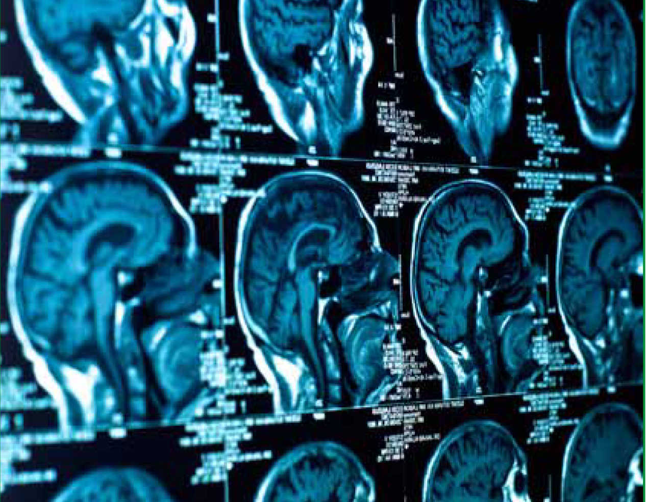
Despite the initial presentation indicating a severe head injury with a large space occupying lesion and raised intracranial pressure sufficient enough to initiate pupillary changes; a CT scan undertaken shortly after arrival in the ED revealed only a small acute traumatic subarachnoid haemorrhage (SAH) at supracellar and interpeduncular cisterns. These cisterns or ‘openings’ in the subarachnoid space of the brain are created by a separation of the arachnoid and pia mater and contain cerebrospinal fluid (Nolte, 2002) and blood associated with a traumatic SAH frequently collects within these areas. Subsequent, contrast CT and MRI scans revealed evidence of intra-parenchymal bleeding, with multiple cerebral contusions, including a focal lesion on the brain stem, affecting cranial nerve 3 and 7. These contusions resulted in a newly formed left sided facial palsy, and dilatation of the patient's left pupil observed at scene. The patient was discharged from hospital approximately one week later with a persistent unilateral facial weakness. On reflection, recognition and subsequent diagnosis of this most complex case in the pre-hospital arena would have been impossible to predict with certainty in the absence of complex computerised diagnostic tests.
Summary statement
Head injuries varying in aetiology, severity and presentation are commonly encountered by practitioners working in unscheduled emergency care in the pre-hospital arena, and remain as one of the major causes of trauma-related morbidity and mortality in all age groups within the UK. These are classically divided into open, closed, crush or penetrating, and involve primary and secondary injuries. Primary brain injuries are a result of initial injury forces, which cause tissue distortion and destruction in the early post-injury period (Greve and, Zink, 2009:97) These injuries lead to alterations in cell function and propagation of injury through processes such as depolarization, excitotoxicity, disruption of calcium homeostasis, free-radical generation, blood-brain barrier disruption, ischemic injury, oedema formation, and intracranial hypertension. (Greve and Zink, 2009). Many of the clinical signs observed would be recognised by health care professionals, either collectively or in isolation and would be indicative of a moderate to severe TBI in the absence of more sophisticated diagnostic imaging, possibly requiring the need for urgent neurosurgical intervention. As such, the decision to undertake an RSI was indicated in accordance with AAGBI and BTF Guidelines, together with our parent organisational Clinical Standard Operating Procedures.
The management of this and other agitated head injured patients continues to pose a real challenge to practitioners operating in the pre-hospital arena. The reduced availability of clinicians competent and experienced in undertaking RSI and procedural sedation, combined with the absence of diagnostic imaging to tailor care specifically to the needs of the patient makes diagnosis extremely difficult. Based largely on the working diagnosis of a TBI, the treatment administered to this casualty adhered to the current best practice guidelines, according to the AAGBI, BTF and parent organisational clinical standard operating procedures. However, limitations in knowledge, skill availability and the limited resources in the pre-hospital arena must be recognised, which often adversely impact on patient care delivery. Despite studies suggesting that drug assisted intubation and RSI undertaken in the pre-hospital arena is associated with increased mortality; it could be argued through clinical experience that RSI when undertaken in accordance with best practice guidelines, remains safe within organisations that operate within regional trauma networks, and where practitioners are comprehensively educated, trained and potential complications are frequently skill-drilled.

Clinical governance processes, including clinical case reviews, robust standard operating procedures, adverse incident reporting, regular appraisals and ongoing audit must all remain integral features in any organisation undertaking controversial interventions. However, RSI is a potentially invaluable skill and an essential asset in the armoury for pre-hospital practitioners to facilitate management of some of the most challenging clinical incidents infrequently presented to pre-hospital practitioners, in an uncertain, unpredictable and often hostile environment. This area of pre-hospital anaesthetic care provision requires significant initial and ongoing investment in training and education, specifically in relation to applied pharmacology and complex decision making skills which are fundamental to the effective assessment and management of critically injured patients. This notion further reinforces the concept of paramedic independent prescribers to improve early access to vital medications in the quest to deliver optimal patient care. However, implementation would require significant organisational and cultural changes, which may further contribute to addressing the lack of pre-hospital practitioners able to safely perform RSI, so that more patients who become critically ill, or injured and who require this clinical intervention, actually receive it in a timelier manner.
‘Head injuries are classically divided into open, closed, crush or penetrating, and involve primary and secondary injuries.’
