
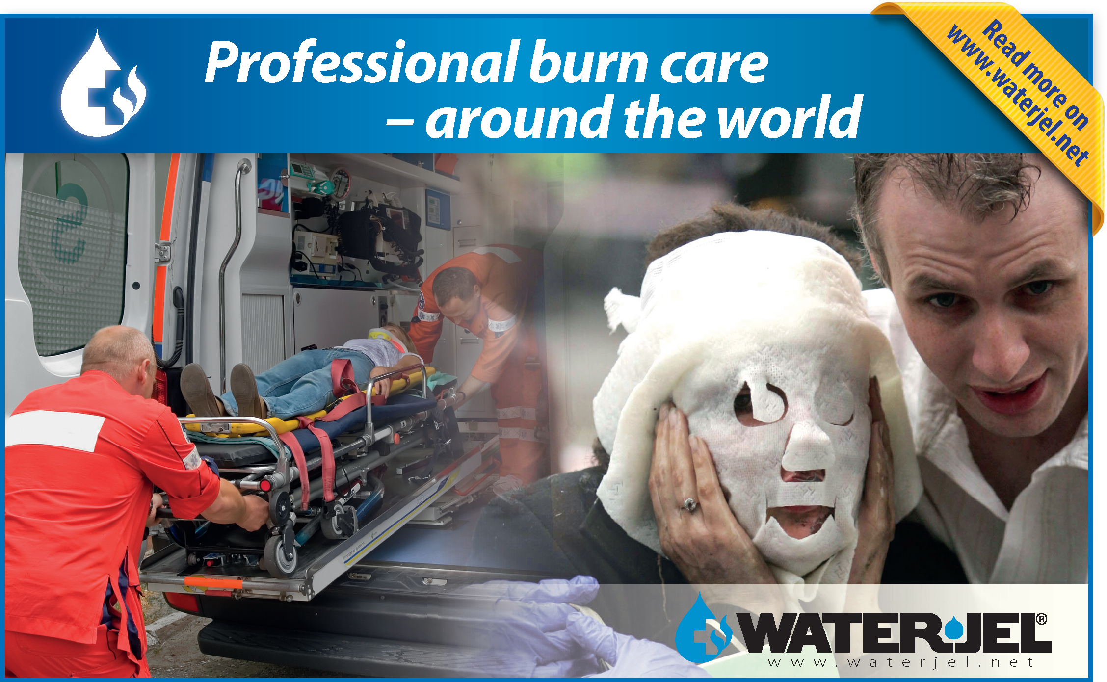
Essex and Herts Air Ambulance Trust (EHAAT) is a publicly funded charity that operates two doctor-paramedic pre-hospital care teams to provide support to land ambulance crews in two counties in the South East of England. The service is predominantly helicopter-based but operates a rapid response car outside daylight hours or in poor weather. The total population covered numbers approximately 3 million people. The team responds to major trauma and medical emergencies, with the latter accounting for approximately 20% of all missions. The paramedics who work for the service are all employed by the East of England Ambulance Service NHS Trust, and are seconded to EHAAT for a period of 24 months. A comprehensive selection process is undertaken and an extensive training programme covering aviation practice and extended clinical management is in place for successful candidates. In addition, EHAAT paramedics have been among the first to enrol and undergo enhanced skill training in anaesthetics and intensive care medicine as part of a postgraduate certificate in advanced paramedic practice in critical care. We believe that one of the strengths of the service provided by EHAAT lies in our paramedics acting as role models and ambassadors for both our service and the ambulance service in general.
The case
We report the case of a 2-year-old boy who sustained a scald to the abdomen after pulling a freshly made cup of tea over himself. Following a crew request for analgesia and transfer to a burns unit, the EHAAT team were mobilised to scene. On arrival the patient was in the back of the ground ambulance, in his mothers arms. She reported that the child had been wearing two items of clothing on his upper body when he pulled a cup of freshly made but milky tea onto his torso, approximately 30 minutes prior to our arrival. The clothing was immediately removed and the scald cooled in the sink with cold running water until the first paramedic arrived. A gel-based burns dressing (Water-Jel, Water-Jel Europe LLP, Hertford, United Kingdom) was applied to the child's abdomen, which was then covered in plastic wrap. On examination, the child was alert, well perfused, crying and moving all four limbs. Given the child's distressed state, it was decided that analgesia was the most important intervention and an estimated weight-based dose of oral paracetamol was administered, followed by one dose of intranasal diamorphine, provided by the EHAAT team. Following analgesia the child was considerably more co-operative and appeared comfortable. The initial dressings were removed in order to assess the extent of the burn. Erythema and some areas of blistering were noted. It was estimated that the blistered areas were substantially less than 10% of the total body surface area (TBSA) and that no ‘special areas’ were involved (Table 1). Due to the short distance the child was transferred by land ambulance to the local district general hospital, accompanied by the EHAAT team due to the administration of intranasal diamorphine. The child was discharged later that day with a follow up appointment for dressings clinic four days later.
|
|
Special areas—areas of the body that if affected may require specialist input for the best cosmetic and functional outcome regardless of depth or size of burn
Discussion
A burn is a traumatic injury to the skin in which the cells of the skin, and in some cases, the underlying tissues are damaged by heat, cold, electricity, radiation or chemicals. Burns and scalds are a form of heat burn that are relatively common in children and can be distressing for all involved. They often occur around the home in seemingly innocuous circumstances, but may also be a result of non-accidental injury. Having the potential to cause death, long life disfigurement, and dysfunction from scarring, an expeditious, systematic approach to paediatric burns is essential to ensure the best possible outcome.
In the pre-hospital setting, the priorities for the management of patients with a burn are to identify immediately life-threatening effects (such as airway compromise, inhalational injury, or hypovolaemic shock), limit the progression of the burn through cooling, and to manage pain in order to perform a thorough assessment of burn size and depth, and to improve the patient's experience. Airway and breathing complications will not be discussed further. The rest of this discussion will focus on the assessment and management of the burn itself, analgesia options, and factors contributing to triage decisions.
Cooling
Effective cooling of a burn can be achieved with simple application of cool running tap water (Venter et al, 2007). Ice and ice-cold water may lead to tissue injury and must not be applied. The timeframe of 20 minutes of cooling advised by the Joint Royal Colleges Ambulance Service Liaison Committee weighs a period of effective intradermal cooling against the potential for induction of hypothermia with prolonged cooling and evaporative heat loss through a disrupted dermal barrier (Joint Royal Colleges Ambulance Liaison Committee, 2013). Large surface area burns compromise the ability of the skin to regulate temperature and retain moisture, which in paediatrics is even more prevalent. Hydrogel dressings, such as Water-Jel and Burnshield (Levtrade International, Johannesburg, South Africa) are designed to cool wounds and provide analgesia in the pre-hospital setting. The results of a study by Coats et al testing different methods of use of a Water-Jel dressing suggested that the same considerations may apply with hypothermia and prolonged use for larger burns unless the dressings are covered as per manufacturer's guidance (Coats et al, 2002). The covering process maintains skin temperature after initial cooling and if the dressing covers a large area, the patient may be exposed to the risk of excess heat loss and hypothermia. We suggest that a practical approach would be application of a hydrogel dressing for burns that are <10% TBSA to provide cooling and prolonged analgesia. For larger burns (over 10% BSA), we suggest that the dressing is covered after application and that continuing pain is assessed and managed appropriately with enteral and/or parenteral analgesic medications. These latter recommendations are consistent with findings from the study outlined above and with personal experience of the authors in the pre-hospital and emergency department setting.
Following cooling of the burn with water, application of plastic wrap (polyvinylchloride film) may theoretically provide some pain relief through prevention of air movement across the disrupted dermis of deeper burns. There may also be some benefit in terms of providing a sterile temporary barrier against infection. Circumferential application of cling film must be avoided because damaged tissues can become oedematous, the swelling restricted by the cling film, and distal blood flow reduced. The authors advocate this simple method of analgesia for non-chemical burns if a dressing that is specifically designed for the purpose is unavailable. Chemical burns should be thoroughly irrigated for a minimum of 15 minutes and until a decrease in pain occurs. In the case of powder chemical burns, safely brush the visible powder away before commencing irrigation. A simple saline-soaked dressing can be used to cover the chemical wound after irrigation, or individual manufacturers for use also licenses some gel-based dressings after chemical burns.
Assessment
Burns are described in terms of depth and an estimation of the size of area that is involved. The size of a burn is determined as a percentage of the total body surface area (TBSA) that has been damaged, excluding any areas of simple erythema. There are a number of tools that can assist in the estimation of the size of a burn; the ‘rule of nines’ may be appropriate for an older child (Hettiaratchy and Papini, 2004), and paper-based diagrams or electronic device applications may incorporate a method such as the Lund and Browder chart (Lund and Browder, 1944). Another well-described method is that of using the patient's hand to estimate the size of the burn (Nagel and Schunk, 1997). Traditionally, the palm of the hand represented 1% of the TBSA; however, more recently it has been described that the palm more accurately represents 0.4% TBSA and the whole hand 0.8%. It is difficult to truly know the size and extent of the burn during the first contact meeting in the pre-hospital environment as a burn matures and develops over the course of 72 hours (Goutos and Tyler, 2013). Therefore, in the pre-hospital phase of care, it must be recognised that descriptions form an estimate only, that a 1% estimation using the extended hand is reasonable, and that the function of this estimate is to guide initial fluid therapy and, to a certain extent, triage decisions.
The depth of the burn is related to the thickness of the skin, the temperature of the heat source and the duration that the heat source is in contact with the skin. Burns are classified as superficial, superficial partial thickness, deep partial thickness, and full thickness. A summary of the features of burns of each depth is included in Table 2. Superficial burns are red erythematous areas of injured tissue that are painful and blanch with pressure. Often, no specific medical care is necessary as they usually heal within two weeks, similarly to a healing area of ‘sunburn’. Partial thickness burns extend through the epidermis and into the dermis. These types of wounds are often very painful due to the exposure of nerve endings. Superficial partial thickness burns are often pink or red and involve blistering of the skin that appears moist, and weepy with serous fluid. They may heal spontaneously over a period of 2–3 weeks following local wound care. Deep partial thickness burns are identified by unroofed blisters, that typically have a white or yellow waxy appearance. In addition, deep partial thickness burns do not blanch with pressure. Although these burns may heal spontaneously over 3–6 weeks, surgical debridement may be necessary to remove devitalised tissue in addition to allowing accurate wound assessment. As the depth of the burn increases, nerve endings are destroyed and therefore full thickness burns are invariably insensate. As the whole of the dermis is destroyed they appear white, leathery, constricted and inelastic. A very small full thickness burn can heal spontaneously but these deeper wounds often require excision and subsequent grafting (Hettiaratchy and Papini, 2004a; 2004b). Burns that extend into muscle or bone are severe injuries are termed ‘fourth degree’ burns, but are rarely encountered.
| Appearance | Clinical features | Typical treatment | Prognosis | |
|---|---|---|---|---|
| Superficial | Red skin, ‘sunburn’ | Mild discomfort, mild swelling | Simple moisturising creams and analgesia | Hospital treatment not usually required |
| Superficial partial thickness | Blistering or obvious areas of superficial skin peeling with ‘weeping’ | Very painful, blanches with pressure, sensitive to light touch | Debridement of superficial dead tissue, analgesia according to level of pain, specific dressings | Usually assessed in hospital and followed up in out-patient setting for wound review and dressing changes |
| Deep partial thickness | Visibly deep wound, appears white | Remains painful and sensitive to light touch (but less than superficial form). Blanches with pressure but return of colour | Often requires wound excision and grafting. Smaller areas may heal depending on vascular supply | Several weeks to heal. Likely to scar and remain light-sensitive for many years |
| Full thickness | Dry, leathery, charred wound without blisters | Insensate, no blanching with pressure | Usually requires excision and grafting by a burns specialist | Usually will not heal without surgical intervention |
| Fourth degree | Obvious extensive injury with muscle and bone visible | Associated with other injuries and physiological disturbance | Requires hospital admission, may require extenseve surgical intervention and grafting with critical care admission | Likely to be associated with poor functional outcome. Increased mortality from effects of burn and associated injuries |
Fluid therapy
Fluid loss from burns may be extensive and as such early fluid management is necessary in major burns to maintain perfusion of vital organs and ensure tissue oxygen delivery. In children this is widely accepted to apply to of burns of greater than 10%. Children become hypovolaemic from burns because of the breakdown of the normal fluid-retaining barrier created by intact skin, and because of the extensive tissue oedema that can occur with large burns.
The local area surrounding the burn wound can be described in terms of three zones (Goutos and Tyler, 2013). The zone of coagulation forms the centre of the burn wound. Here, cells have suffered the most intense heat and are damaged beyond repair. These cells will be unable to regenerate and will require debridement. The zone of stasis encases the zone of coagulation. This area of tissue is intensely fragile, suffering from microcirculatory ischaemia due to fluid loss and oedema. The outermost zone is called the zone of hyperaemia. Within this zone, minimally damaged cells are able to spontaneously regenerate over a short period of time. One of the aims of fluid resuscitation is to maintain perfusion to the salvageable watershed area of the zone of stasis.
The Parkland formula is a common fluid management programme that takes into account millilitres of fluid per kilogram, per percentage TBSA burned (Baker et al, 2007). Half of the total amount is provided within the first 8 hours following burn injury, with the other 50% administered over the next 16 hours. The 8 hour timeframe to administer the first portion of fluid begins at the time of the burn. If the journey time to the hospital is relatively short (less than 30 minutes) and the patient is not shocked, it may be suitable to wait until arrival in hospital for fluid resuscitation to be commenced with specific physiological targets to guide therapy. The physiological goal in the pre-hospital arena is to ensure that cerebral perfusion and vascular pressure is maintained in order to meet the metabolic demand of the tissues, therefore improving the survival of cells within the zone of stasis. Guidance for paramedics in the United Kingdom recommends administering bolus doses of intravenous crystalloid using a 10 ml/kg-dosing regime (Joint Royal Colleges Ambulance Liaison Committee, 2013).
Analgesia
Pain management in burns patients should be multimodal. Early administration of enteral and parenteral analgesia aids in calming the child and therefore improves their experience. Parents should be involved early on in the management process to assist in comforting the child. Much of the initial assessment can be performed from afar until the child's trust has been gained and distress lessened. It is helpful to use a recognised method of objective pain scoring to guide analgesia provision (Atkinson et al, 2009). In alert children, weight-based doses of paracetamol, ibuprofen, and oral morphine solution (guided by the objective measure of pain) will provide initial analgesia with moderately quick onset. Immediate analgesia involves the administration of appropriate weight-based intravenous boluses of opiates such as morphine. Some services and enhanced care teams may have access to intranasal preparations of diamorphine, fentanyl, or midazolam. This route is useful for rapid administration of analgesia but is better considered a bridge to gaining intravenous access and correctly titrating intravenous analgesia (Atkinson et al, 2009). Ketamine is often used in the hospital setting for analgesia and may be available in the pre-hospital setting for enhanced care practitioners. Delivery of a general anaesthetic to a child solely for pain control in the context of burns is rare and is a complex decision that is outside of the scope of this article.
Triage
Triage to hospital is a decision that is made in the context of what a specialist burns centre can offer that is not available at a non-specialist local hospital. Ultimately, local policy will dictate which cases warrant direct triage to a burns centre, and local protocols and agreements will be in place to empower clinicians to take such action. The National Network for Burns Care lists a minimum threshold criteria of injuries requiring referral to a burns care service, but the majority of these do not necessitate direct transfer from the pre-hospital environment (National Network for Burn Care, 2012). Many specialist burn care services now provide comprehensive telephone advice for other hospitals during the initial stages of care, in addition to facilitating interfacility transfers as required and providing dressing clinics later in the care pathway. Therefore, it is appropriate in many cases to transfer the patient to the nearest hospital and if necessary, for them to be transferred to a specialist centre further on in their care pathway. Certainly, the initial management of even extensive burns can be dealt with in most emergency departments in the United Kingdom. Enhanced care teams, in line with pre-agreed protocols and criteria, may take patients directly to a specialist burns centre on a case-by-case basis.
The role of enhanced care teams
The majority of burns can be managed effectively with detailed attention to the aspects of pre-hospital care outlined above. There are a small number of cases for which an enhanced medical care team, such as the EHAAT critical care team may be able to offer additional value. Specific examples may be cases in which there has been concomitant major injury, cases in which airway obstruction has occurred or is imminent, and cases in which it is suspected that the patient is unconscious from inhalational injury such as carbon monoxide or cyanide poisoning. Helicopter-based teams may be able to assist with the transfer of suitable patients over large distances to a burns unit, especially if there is room for a parent to travel too.
Conclusions
We have highlighted the need for early adequate analgesia, thorough assessment, and triage to the correct hospital where the patient's injury can be appropriately managed. While the majority of burns can be expertly managed in the pre-hospital setting using basic techniques, we have also highlighted areas in which an extended care service such as that provided by EHAAT can help, particularly in terms of supplementary analgesia and transfer to specialist units.