The ability to secure a patient's airway is crucial in the management of acutely life-threatening illnesses and injuries (Mulryan, 2011). Endotracheal (ET) intubation can be hazardous, particularly as patients may have deteriorated rapidly or have combined respiratory and cardiovascular failure. These forms of emergency situations are frequently encountered by paramedics, necessitating an acute awareness of their role in emergency airway maintenance.
Although supplementary oxygen can improve oxygenation and oropharyngeal airways can help maintain a patent airway, both require that the patient spontaneously breathes. However, when cessation of breathing occurs or when respiratory rate and effort is insufficient to maintain normal respiratory function, ET intubation may be required. Paramedics are required to competently achieve ET intubation in often time-critical situations, in less-than-ideal conditions.
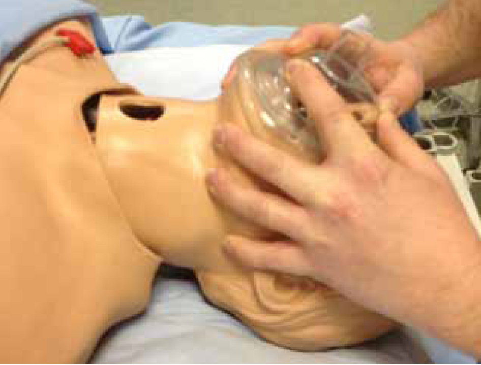
Initially, in an emergency situation if simple techniques are not successful a bag valve mask may be the most effective way to assist ventilation. Paramedics need to be able to use this equipment by ensuring a tight seal is produced at the patient's face. This is done by using jaw thrust or maintaining the head tilt manoeuvre. Castle (1999) suggests that a two-person technique provides more effective ventilation for the patient. The success of this practice is dependent on the skill of the practitioner, as poor techniques could hamper efforts to maintain the airway. The airway should be cleared of foreign substances, such as sputum or vomit, through the use of suction. In the majority of cases the patient's airway is occluded by relaxation of pharyngeal and jaw muscles, causing tissue to dramatically narrow the airway and the tongue to fall backwards over the larynx. Therefore, by thrusting the mandible/jaw forwards, the tongue will be lifted away from the larynx, thus clearing a path for ventilation. Care should be taken when performing this manoeuvre on elderly patients, as pressure on soft tissues below the mandible during jaw thrust could inadvertently occlude the airway. This may be identified by visualising finger positions and any difficulty in ventilating (Figure 1).
False teeth can both aid and hinder bag and mask ventilation. If false teeth are securely fitted they can help to maintain the shape of the face, thus facilitating a good seal with the mask. Conversely, if the false teeth are loose they could contribute to airway occlusion, in which case they should be removed (Allman et al, 2009).
Several other factors have been identified as predictors of difficult bag and mask ventilation (Walls et al, 2011). Kheterpal et al (2006) maintain that one such factor includes obesity (BMI >30 kg/m2) and a history of snoring, as these indicate the mandible will freely fall backwards and of course would be heavy and potentially difficult to thrust forwards. Forward excursion of the mandible can be facilitated by using a two-person technique, with one practitioner performing jaw thrust, while the other ventilates; the addition of an oropharyngeal airway could further aid ventilation attempts. As the mask seal requires a good contact between the mask gasket and patient's skin, the presence of a beard can result in leaks and inadequate ventilation. Allman et al (2009) suggests sticking a transparent dressing over the beard with a hole cut to allow for ventilation of the mouth and nose, which can somewhat alleviate this issue.
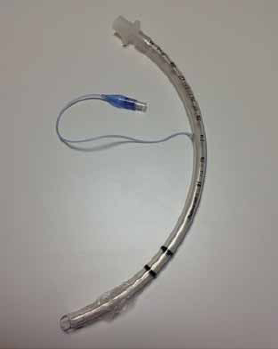
Once an airway is established the bag valve mask allows for greater amounts of inspired oxygen to be delivered through the supplementary oxygen and the reservoir bag used with the system. When paramedics use this system they should observe for adequate chest expansion during inspiration, avoiding over expansion as this may lead to lung injury. Care should be taken to allow for expiration to occur in order to ensure carbon dioxide expulsion. However, this means of ventilating a patient is generally used for short-term purposes. If ventilation is required for prolonged periods in an emergency situation, then ET intubation should be performed. This involves the placement of a cuffed endotracheal tube (ETT) in the trachea (Figure 2).
Endotracheal tube placement
Ideally, the placement of an endotracheal tube should be performed in a controlled environment, although this is rarely possible in a community setting. With the high probability of gastric contents being present in the emergency patient, a rapid sequence induction (RSI) is desirable to minimise potential for pulmonary aspiration. This requires two practitioners, one to apply cricoid pressure, which involves placing the thumb and index finger on the cricoid cartilage, which lies inferior to the thyroid cartilage. The objective of this pressure is to compress the oesophagus between the cricoid cartilage and the C6 vertebrae. In theory, this will dramatically reduce the risk of gastric aspiration during intubation; however, some studies have suggested this techniques does not confer safety if best practice is not maintained (Thorn et al, 2005; Gudzenko et al, 2010). The pressure exerted should be 30 Newtons (N); this can be practiced by filling a 50 ml syringe with air, sealing the syringe with a bung and compressing the plunger to 38 mls (Flucker et al, 2000; Kopka and Robinson 2005). This pressure is equivalent to 30 N, and can be practiced to provide the paramedic with the ‘perceived feel’ of this correct pressure. The paramedic should take note that once cricoid pressure is applied it should not be released until the ETT balloon is inflated and correct placement is confirmed by the intubating paramedic. Therefore, it is vital that all required equipment is at hand and checked for functionality before induction and intubation is attempted, including bougies and laryngeal mask airways (LMAs) in case of difficult intubation (Jaber et al, 2010). An algorithm provided by the Difficult Airway Society (Henderson et al, 2004) can offer guidance for the team when attempting RSI and appropriate actions if difficulties are encountered. The inflation pressure of the ETT balloon should not exceed 15–25 mmHg; however, initially the balloon should be inflated enough to provide a seal thus protecting the airway, with adjustments to the balloon pressure made immediately after ETT placement confirmation to avoid tracheal necrosis (Galinski et al, 2006). Table 1 outlines essential equipment for endotracheal intubation.
| Laryngoscopes x 2 (3 & 4 MAC Blade) (e.g. Portex Standard Light Disposable Laryngoscope Blade; Visionary® Classic Laryngoscope; Visionary®! Fibre Optic Laryngoscope; PROACT Metal Max™) |
| Endotracheal tube (sizes 6,7,8 & 9) (e.g. Intersurgical InTube cuffed and Uncuffed ET Tube; Portex Clear PVC, Oral/Nasal, Soft Seal® Cuff Tracheal Tube; Portex Siliconised PVC, Oral/Nasal Uncuffed Tracheal Tube) |
| 10 ml Syringe |
| Lubricating gel (aqueous gel) |
| Bougie (e.g. Portex ET Tube Introducer) |
| Laryngeal mask airway (size 3,4 & 5) (e.g. LMA Intravent Laryngeal Mask Airway; Ambu laryngeal mask; Portex Single-Use Soft Seal Laryngeal Mask) |
| Macgills forceps |
| Oropharyngeal airway (sizes 2,3 & 4) (e.g. Intersurgical One-piece Guedel Airway) |
| Securing tapes |
| Endotracheal tube cuff pressure manometer |
| Suction with yankauer sucker (e.g. Portex SuctionPro 72™) |
However, in a time-critical emergency situation, the paramedic's primary objective should be to conserve life through achieving a definitive airway, as rapid response paramedics would not have the addition of a second practitioner to provide cricoid pressure. Therefore, RSI may be an unrealistic option in these cases, as hypoxia will kill quicker than aspiration, which is treatable to some degree.
Endotracheal intubation
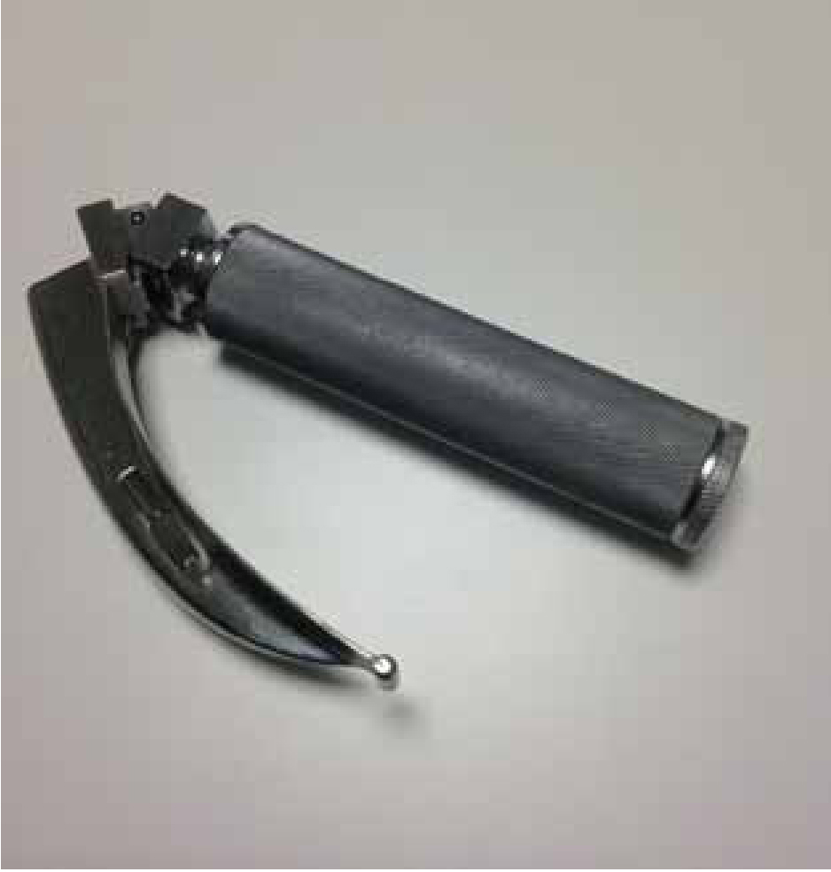
Tracheal intubation has multiple potential problems that must be anticipated in order for adverse events to be avoided (Martin et al, 2011). Each tracheal intubation event should be anticipated as a potentially difficult intubation. Although airway assessment methods such as the Mallampati scoring system are available, they can be difficult to carry out in acute emergency situations and their effectiveness has not been evaluated in emergencies (Orebaugh, 2002). Once the patient has been sedated or is deeply unconscious, the process of intubating can begin (Woodall et al, 2011). This is achieved through the use of a laryngoscope, there are varying types of blades available, but generally a Macintosh (MAC)-style blade is used (Figure 3).
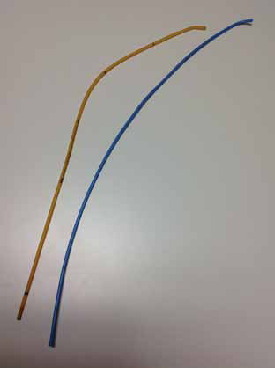
This blade enables the intubating paramedic to lift the tongue and mandible forwards, which along with ‘head tilt’ should provide a clear view of the larynx. There are instances, however, when a patient's larynx may be anteriorly situated, thus making intubation difficult. This can be overcome by applying pressure to the larynx and pushing it backwards, upwards and to the right, termed the BURP manoeuvre (Allman et al, 2009). However, any external manipulation of the larynx should be under the intubating paramedic's guidance in order to improve laryngeal view (Berkow, 2004). These manoeuvres may bring the larynx into view enabling the passing of the ETT into the trachea. Additional techniques could be the use of a bougie (Figure 4), this is passed into the trachea and the ETT threaded over the bougie, guiding the tube into the trachea. Following loss of consciousness, the protective airway reflexes of cough, gag and swallow are lost, meaning there may well be bodily fluids pooling in the airway. If these fluids gather on the vocal cords they could instigate laryngospasm, whereby the larynx close during inspiration in order to protect the lungs from aspiration. This protective reflex paradoxically makes ventilation extremely difficult, it is therefore imperative that any bodily fluids are cleared promptly from the airway using suction with a yankauer sucker. The paramedic should be cautious, however, to avoid causing damage to delicate airway tissues with the yankauer sucker, as this could cause bleeding or tissue oedema, both impairing airway patency.
If these techniques fail to achieve tracheal intubation then an LMA could be inserted and in most cases (~95%) this will enable oxygenation (Allman et al, 2009) until a more definitive airway, usually a cricothyroidotomy, can be established.
The Advanced Life Support Manual (Resuscitation Council UK, 2011) suggests the usual size ET tube required for male ET intubation is 8–9cm and for women 7–8cm. The oral ET tube should be positioned at about 23 cm at the incisors for men and 21 cm for women (Nseir et al, 2011). The tube should be positioned about 2 cm above the carina, which can initially be assessed by observing for bilateral chest rise on ventilation and auscultating all lung zones for air entry. Once the paramedic is satisfied the tube is correctly placed he/she must note the position of the tube at the patient's incisors using the graduations on the tube.
Of course, it is of great importance to secure the ETT once it is in place to prevent it from moving. This can be achieved through the use of ETT ribbon or specially-manufactured devices. These are secured to the ETT and then passed around the patients head, then tied sufficiently to offer secure fastening for the ETT, but not so tight as to cause pressure ulcers, which tend to develop at the side of the patient's mouth or around the occiput. In cases where the patient has a suspected head injury, the ETT should be secured with tape to the cheek bones and mandible. This method ensures maintenance of cerebral venous drainage, avoiding the development of cerebral oedema from the ETT securing technique. Whatever securing technique is employed, the paramedic needs to regularly note the tube position by observing the graduations on the tube itself at the patient's incisors. This is particularly important in paramedic practice as movement of the patient onto a backboard and/or stretcher provides the potential for tube displacement, leading to ineffective ventilation. If suspicions arise that the ETT has moved, this should be immediately acted upon and the tube pushed back into the original position or reintubation undertaken if the tube is pulled beyond the cords. Factors that could lead to such suspicions are unilateral chest rise on ventilation, difficulty in ventilating and/or reducing oxygen saturations. In addition to these factors, ventilation and oxygenation should be assessed through pulse oximetry and arterial blood gases, with any problems addressed immediately.
The indications for intubation usually means that the patient is respiratory compromised, has little or no spontaneous respiratory effort, is unable to maintain their own airway, and is unable to remove CO2 and/or maintain adequate levels of O2 in the arterial blood (Finucane et al, 2011). Once a patient has been intubated, the respiratory function will need to be facilitated through the ETT and often this is undertaken via an oxylog ventilator for conveyance to hospital. The use of a ventilator enables consistent and controlled ventilation, reducing the risk of lung hyperinflation and control of FiO2 delivery. Invasive ET intubation is the ‘gold standard’ of airway management (Oh et al, 2008), providing airway protection and a route for positive-pressure ventilation.
Mechanical ventilation
Mechanical ventilators are designed to assist the movement of gases (air) into and out of a patient's lungs while minimising the work and effort of breathing (Scholz et al, 2011). Most mechanical ventilators used today within the UK are positive pressure ventilators, where lung volumes and gas exchange are achieved by applying a positive pressure to the thorax (through the trachea), via an ETT.
There are many different types of positive pressure ventilation strategies and ventilator modes. Essentially, ventilators deliver gas by employing a controlled-cycling mechanism, such as a pressure-cycled, time-cycled or volume cycled mechanism. Pressure-cycled strategies occur when a volume of gas is delivered until a pre-set pressure is reached; time-cycled strategies involve the delivery of a volume of gas until a pre-set time is reached; and volume-cycled strategies involve the delivery of a volume of gas until a pre-set tidal volume is reached.
The maintenance of adequate tidal volume is important in maintaining normocapnia, with recommended tidal volumes being 6–7 ml/kg (Singer and Webb, 2009). Respiratory rate can also be set on the ventilator, the objective of setting both tidal volume and respiratory rate is to maintain a minute volume, which can be expressed as follows:

Failure to maintain normocapnia can lead to acidosis, which has depressive effects on cardiac contraction and decreases vascular tone, leading to haemodynamic instability.
It is beyond the scope of this paper to discuss all ventilator strategies and ventilator modes; therefore, Table 2 provides an outline of those that are commonly used.
| IPPV = Intermittent positive pressure ventilation | This mode of mechanical ventilation is used to deliver pre-set inspiratory pressures to achieve the desired tidal volume. Used in patients with no spontaneous breathing. |
| SIMV = Synchronised intermittent mandatory ventilation | Allows the ventilator to detect a patient's own breathing and permit spontaneous breathing between ventilator breaths. Also provides ventilator assistance to a patient's own breath. Used to facilitate weaning from mechanical ventilation. |
| BiPAP = Biphasic positive airway pressure | BiPAP combines pressure-controlled ventilation with unrestricted spontaneous breathing. BiPAP ventilates using time-cycled switching between two pressure levels: Phigh and Plow, with switching being determined by preset time intervals. The volume displacement caused by the difference between the two pressures levels and the rate at which these changes occur contribute the mechanical ventilation. At both pressure levels a patient can breathe spontaneously in a continuous positive airway pressure system (CPAP). |
| CPAP = Continuous positive airway pressure | CPAP delivers a continuous positive air pressure throughout the respiratory cycle in spontaneously breathing patients. |
| PEEP = Positive end-expiratory pressure | Not an actual ventilator mode but an important respiratory concept: The patient's airway is kept open above atmospheric pressure at the end of expiration. |
Suctioning
If a patient has an ETT in situ, it might be necessary for the paramedic to perform endotracheal suctioning. This is done to remove secretions from the airways and it is another essential skill that paramedics need to be able to perform effectively. ET suctioning can improve the patency of the airway, improve oxygenation and improve gaseous exchange (Viney, 1999).
To perform ET suctioning appropriately, the correct size suction catheter should be used (Figure 5). In order to work out the correct size of the catheter for suctioning, the following formula should be used:

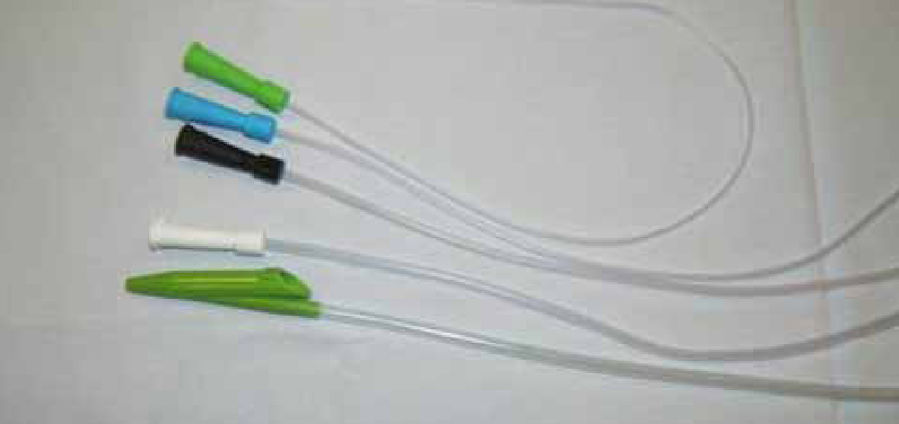
For example, a patient with a size 8 ETT will require a size 12 suction catheter.
Hypersalivation also occurs when an ETT is in situ, therefore the paramedic must ensure that excess saliva is cleared from the patient's mouth and oropharyngeal area. Furthermore, a patient will not have the active airway protective reflexes of cough, gag and swallow once anaesthetised or sedated. Consideration should also be given to the fact the oesophageal sphincter relaxes during anaesthetic induction, which, when coupled with the routine insertion of a nasogastric tube means there is a great risk of gastric aspiration. These bodily fluids could potentially bypass the ETT and enter the lungs, causing aspiration pneumonia, with ~25 mls of gastric fluid associated with high rates of morbidity and mortality (Patten, 2006). Thus regular oropharyngeal suction and high standards of oral care are essential to reduce risk.
Conclusions
Maintaining a patient's airway and facilitating breathing are the main priorities in any emergency situation in which breathing is compromised. This article has provided paramedics with an overview of equipment and techniques regarding airway management and ventilatory support that may occur within an emergency situation in which they are involved. The emergency techniques of supplemental oxygen therapy and insertion of an oropharyngeal airway tube are briefly discussed before more detailed consideration of the techniques, benefits and risks associated with bag-valve-mask ventilation, and endotracheal tube management, mechanical ventilation and endotracheal suction, which is required for safe removal of respiratory secretions.